Smart Fusion of modalities enhances clinical output
Adding high quality, dynamic ultrasound for hybrid imaging enables clinicians to improve detection of a range of lesions or to intervene better for improved clinical outcomes. ‘We can no longer be fascinated with pictures; what we need is proof of the clinical benefit from tools and techniques,’ said Professor Jose Zamorano MD, Director of Cardiology at Ramón y Cajal University Hospital in Madrid.
Currently, he is building a multi-centre study of cardiovascular imaging (CVI) to evaluate the severity of ischemia using a hybrid display combining CT Angiography and 3-D ultrasound called Smart Fusion from Toshiba.
Meanwhile, at the University of Paris Necker Hospital, Jean-Michel Correas MD has also moved beyond fascination with the advanced capabilities of Smart Fusion by applying the technology to clinical practice. He has added advanced contrast-enhanced ultrasound images in real-time to both CT and MRI acquisitions better to target and treat even small isoechoic or non-visible lesions. ‘There are clear benefits for lesion detection, Dr Correas said, adding, ‘as well as for treatment planning with the possibility of finding new routes to the lesion, which is a key advance.’
At the University of Berlin’s Charité Hospital, Thomas Fischer MD, Director of the Ultrasound Research Lab Radiology and Head of Ultrasound Diagnostics at the Institute of Radiology, finds the enhanced capabilities of hybrid imaging with Smart Fusion makes navigation more comfortable for positioning and performing ultrasound-guided biopsies of prostate tumours. ‘Not all forms of prostate cancer are created equal,’ he pointed out. ‘Certain patients, those who have a non-aggressive form of prostate cancer, will not benefit in any significant way from the therapy. Thus we need to identify the tumours that will respond to the therapy.’
MRI remains the modality of choice for identifying the exact location of the dominant lesion in the prostate gland. For those patients who will benefit from treatment, the challenge become re-locating the tumour with an ultrasound system for guidance. ‘Here the B-mode image, which is crucial for biopsy and treatment purposes, comes in,’ he explained. ‘Planes from both imaging modalities need to be fused as precisely as possible in order for the biopsy needle to hit the lesion. And this in turn means that I do need to see the needle.’
For this, the Smart Fusion platform offers superior image quality thanks to a special transducer developed by Toshiba. Fusing this high quality dynamic image with the static MRI acquisition of the organ is simplified with a sensor placed on the transducer handle. ‘That’s the trick behind Smart Fusion,’ he explained. ‘This sensor tells the system exactly where the transducer is positioned within the cavity. In other words, the hand movement can be quickly translated on the moving images.’
According to Dr Correas, the combination of dynamic tracking with the new transducer and the image quality effectively creates a new tool for interventional procedures. ‘There is improved lesion detection and characterisation, which is the key step,’ he said, adding, ‘After all, if you cannot see the tumour, it will become very difficult to treat it.
‘Without going into detail about the procedure itself, I can say it is rapid where volume data from CT or MRI were loaded to the Aplio 500 system to allow simultaneous dynamic display. We fix one transverse plane, locate one target point and can then fuse the images.’
The result is a clear benefit for better diagnosis, planning and treatment, ‘…and we can evaluate results almost immediately after ablation to be sure of correct coverage,’ he said.
With a significant patient workload, the reduction in procedure time becomes an important advantage, as well as the benefit of reduced radiation exposure for patients and medical staff.
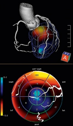
Applying this technological advance to an assessment of cardio-vascular conditions is opening a new field of study for Professor Zamorano in Madrid: ‘Smart Fusion in cardiovascular imaging has the potential to greatly aid in the assessment of coronary artery disease for ischemia and heart failure, an extraordinary advance through the integration of imaging information provided by the CT scan and echo.’
The resulting images display the coronary arteries and branches on a colour-coded myocardial volume, making it possible to correlate the degree of coronary stenosis with the information from myocardial strain in the surrounding myocardial territory. This holds the potential for a non-invasive assessment of myocardial mechanics and the relationship with coronary ischemia, he said. ‘With CT images we get an excellent assessment of the morphology of the coronary arteries, but this is not enough for analysing coronary artery disease, which is caused by functional issues, such as ischemia,’ the professor pointed out.
Where the CT may show a 70% stenosis in the left anterior descending (LAD) artery, he explained, ‘this does not tell you a lot about the ischemia related to that stenosis. What we add with Smart Fusion is the data from stress echo, and now we clearly see if the area of ischemia is related to a specific coronary artery, as well as the severity of the stenosis at that level.
‘We are at the very beginning of the process, and building evidence of the clinical benefit of the CVI technique is very important.’ Prof. Zamorano added that he expects to join with other centres in Europe for a clinical study to validate initial findings. ‘We are a university hospital, very proud of developing a new technique, but ultimately the tools and the technique must be oriented to a real clinical benefit,’ he stressed. ‘So many patients are affected by ischemia, and we are sharply focused on evidence of the clinical outcomes.’
05.11.2013