Will MRI become routine modality? Today, thoracic MRI is rarely performed in Europe. But this will change over the next decade, predicts Professor Hans-Ulrich Kauczor, Medical Director of the Radiology Clinic at University Hospital Heidelberg. He expects Germany to be at the forefront of this development because MRI technology, despite the high costs, is already widely used here and because CT radiation exposure is a big issue in the German healthcare community.
Professor Hans-Ulrich Kauczor
“If we look at Europe as a whole MRI use continues to be mostly a matter of availability. In Slovenia for example there are only one or two MRI systems installed while in Italy the technology is popular and consequently clinical use as well as MR research are well advanced”, Professor Kauczor points out. One Europe-wide trend is obvious: MRI is the modality of choice in paediatrics, for example to study congenital diseases such as cystic fibrosis and heart defects.
Looking at Germany – what will change? “MRI unlike CT does not require radiation exposure, a fact which is of utmost importance for radiation sensitive patients and for repeat exams,” says Professor Kauczor. Thus, he adds, MRI is a very sought after technology whose performance will increase to the point that a few years from now it will be able to replace CT in many routine examinations. Kauczor looks forward to “thoracic MRI being as easy as a knee MR scan: fewer sequences and examination times of no more than 15 minutes.” Consequently, MRI will be used above all with children and adolescents, with women under 40 and with patients who need regularmonitoring, for example patients with cystic fibrosis and heart defects. Ten years from nowthe radiologist is convinced, “staging of lung carcinoma, currently performed with PET-CT, will be done routinely with whole-body MRI.”
Moreover, MRI will complement CT to assess interstitial lung disease since unlike CT MRI can precisely show acute inflammatory processes which is the precondition for any meaningful therapy decision. Nevertheless, Professor Kauczor underlines, “MRI will not replace CT for these patients because the new classifications are predominantly based on morphological imaging, that is CT diagnostics.” MRI will also facilitate evaluation of chronic obstructive pulmonary disease (COPD), he adds: “Complementing conventional pulmonary function diagnostics MRI will help us to classify COPD structurally and functionally and to stage it.” This would for all intents and purposes render CT obsolete in this area, except maybe for a few sub-phenotypes, certain surgery planning procedures and interventional endobronchial and percutaneous therapies.
There is some very promising MR lung research that Professor Kauczor hopes will yield important new insights, particularly different nuclei and MR elastography. In several European countries, xenon and sodium MRI are currently being tested. Research on hyperpolarized xenon may offer new knowledge on lung diseases with reduced oxygen uptake while sodium MRI promises to further our knowledge of homeostasis and breathing mechanics in general. “I consider these research approaches to be very important as they will help us understand many new physiological and pathophysiological aspects“, the expert says; he doubts however that the research will lead to concrete clinical applications.
Speaking about clinical relevance – MR elastography (MRE) of the lung may well be the way to go. MRE is already being used to measure elasticity of liver tissue and might also be suited to measure lung tissue stiffness in patients with cystic fibrosis. This would be a major advancement since currently this kind of lung assessment is rather complicated as Kauczor describes: “Today the only compliance measurement available requires a probe to be inserted in the oesophagus to indirectly measure a single parameter. Regional differences cannot be assessed. Maybe MRE can help us. But that’s still pie in the sky.” We hope to have more concrete news on this pie shortly!
####
Profile
In the late 1980s Professor Kauczor studied medicine in Bonn and Heidelberg and worked as research associate in the radiology department of the German Cancer Research Center – an institution that he would head many years later, from 2003 to 2007. He received his medical degrees from the Universities of Cologne and Mainz. Since 2003 Kauczor has been Professor of Diagnostic Radiology at the University of Heidelberg and in 2008 he was appointed Medical Director of the Radiology Clinic of that institution. In 2000 he was awarded the German Röntgen Society’s Holthusen Ring for his research in radiology. He was ESTI president in 2011.
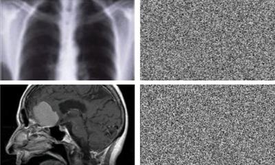
Leveraging the power of chaos theory, experts have developed a new way to encrypt medical images such as X-ray, CT and MRI scans, keeping them secure even if hospital networks are breached.
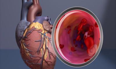
A patient reports with chest pain, but in the coronary angiogram, the main heart arteries look clear, so it cannot be angina – right? A new study reveals that this approach can easily lead to…
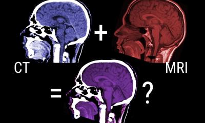
New research shows how AI can be used to fuse images from clinical X-ray CT and MRI scans to allow a clearer and more clinically useful interpretation of the images.
This website uses cookies to give our readers the best website experience. Please refer to our privacy policy to find out how we use cookies and how you can edit your preferences.