When cardiac catherisation delivers no result...
The significant benefits of cardiac catherisation remain undisputed. However, cross-sectional imaging modalities are serious competitors when it comes to arriving at the right diagnosis.
Recently, during the German Radiological Society Congress, Dr Tilman Emrich presented the results of his study on the diagnostic importance of cardiac MRI (CMR) for patients suffering acute chest pain, elevated levels of cardiac enzymes and a negative coronary angiography.
A 23-year-old male without a known pre-existing illness, and a 45-year-old female who had recently suffered a severe blow, were admitted to A&E at Mainz University Hospital and treated in the specialist chest pain department. The ECG showed no abnormalities but blood tests showed elevated troponin levels. Independent of the patients’ age and sex everything pointed towards myocardial infarction. As per the established guidelines for these cases, the patients are therefore taken to the cardiac catheter laboratory. However, the cardiologist could find no evidence of a myocardial infarction.
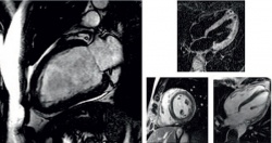
‘In this case, the cardiologist is faced with a dilemma. What should he do – send the patient home or continue treatment without a diagnosis?’ asked Dr Tilman Emrich of the Clinic for Diagnostic and Interventional Radiology at Mainz University Hospital. His answer: ‘In this situation, a heart MRI can be helpful as it enables an examination of the functionality together with the anatomy and analysis of the tissue. The clinical and laboratory results suggested an undiagnosed heart problem for both patients.’
Studies published to date have documented the field of application for cardiac MRI in these cases, although there is as yet no study on a case where a patient’s radiological diagnosis was cross-checked with the cardiologist’s final reference diagnosis in the context of clinical proceedings.
Back in 2007, this prompted Emrich, then still in specialist radiology training, to carry out a cardiac MRI in 125 patients whose cardiac catheter examination did not have any indicatory results, and to compare both diagnoses.
His work was overseen by Professor Karl-Friedrich Kreitner and the study was carried out between 2007 and 2010 - with a satisfactory result. ‘The MRI scan showed multiple cardiac pathologies and in nine out of ten cases the MRI diagnosis concurred with the cardiologist’s final reference diagnosis.
The five most common indications were myocarditis, dilated cardiomyopathy, acute myocardial infarction, Takotsubo cardiomyopathy (Broken-Heart Syndrome) and hypertensive heart disease,’ explains Emrich. The MRI scan helped to make the right diagnosis for all cases of myocardial infarction and Takotsubo cardiomyopathy; in the other cases there were only slight variances.
In the case of four patients, the cardiologists were not able to make a final diagnosis at all.
02.09.2014