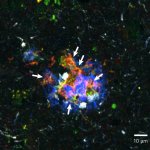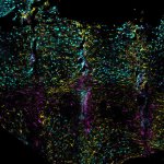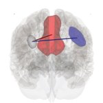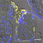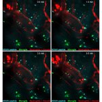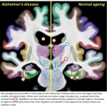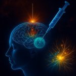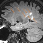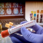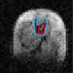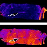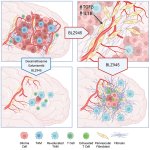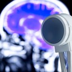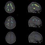
News • OLE-induced microglia rewiring
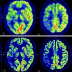
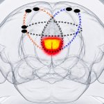
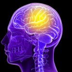
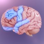
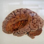
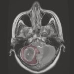
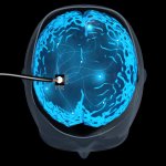
Molecule “reprograms” the brain’s defenses against Alzheimer’s disease
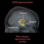
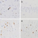
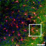
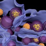
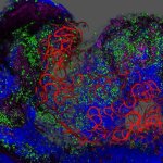
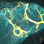
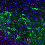
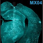
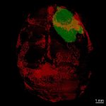
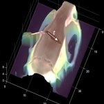
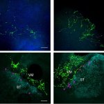
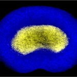
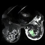
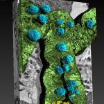
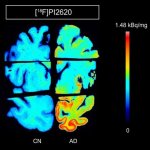
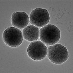
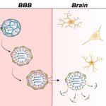
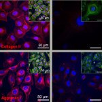
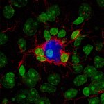
A new experimental compound, called OLE, helps the brain enclose and contain beta-amyloid plaques – a hallmark of Alzheimer’s disease –, reducing their size and toxicity.