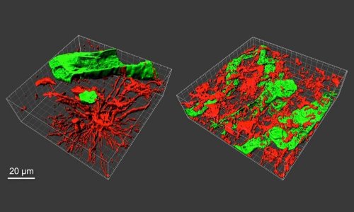
Image source: Andreiuolo, F et al., Acta Neuropathologica Communications 2024 (CC BY 4.0)
News • Neuropathology
Technology offers new insights into pediatric brain tumors
Next-generation molecular sequencing and DNA methylation profile analysis allow the characterization of rare brain tumors
The research results were recently published in the journal Acta Neuropathologica Communications.
Embryonal tumors of the central nervous system (CNS) are a class of malignant tumors composed of embryonal neuroepithelial cells predominantly found in children. Recent updates by the World Health Organization (WHO) classification included several new subtypes of these tumors, highlighting the increasing importance of epigenetic studies in defining these subcategories. Epigenetic studies, particularly DNA methylation profiling, have an increasing importance in the classification of brain tumors and new tumor types delineation. The technique application allowed the identification of a particular group of cases where genetic analysis revealed a specific molecular alteration (fusion involving the BRD4 and LEUTX genes).
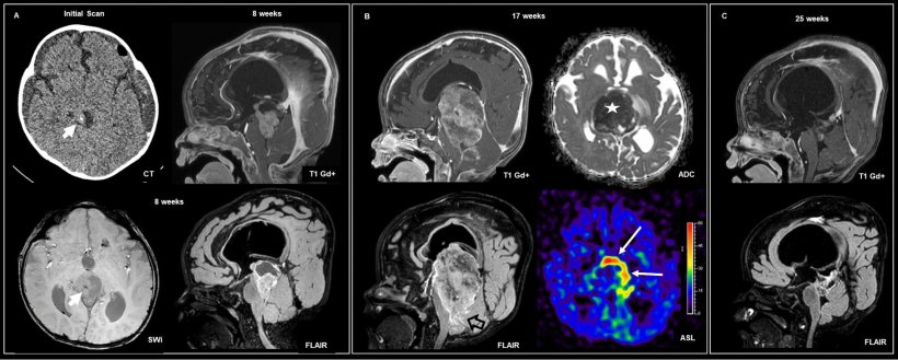
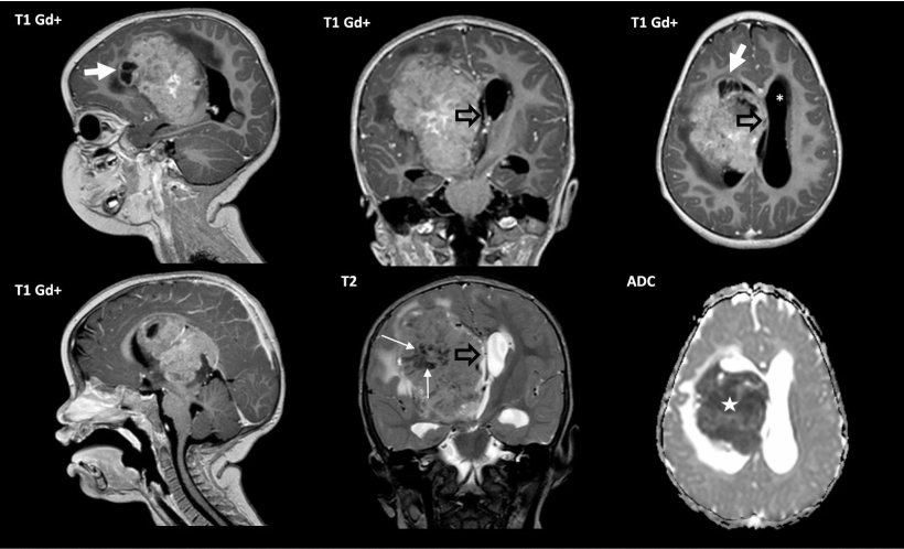
Image source: Andreiuolo, F et al., Acta Neuropathologica Communications 2024 (CC BY 4.0)
The BRD4 gene (Bromodomain Containing 4) acts in transcriptional regulation and development, as well as maintaining the pluripotency of stem cells, and its dysregulation has been associated with different types of cancer, as it contributes to cell proliferation and tumor progression. On the other hand, the LEUTX gene (Leucine Twenty Homeobox) plays a role in embryonic development and regulation of early cell differentiation.
In this study, careful analysis of four new cases with these molecular alterations and five other cases described individually in the literature revealed similar clinical, radiological, and histopathological characteristics, indicating that they are most likely a specific type of tumor. All patients were children up to 4 years old, with bulky tumors, rapid growth, and aggressive clinical behavior. However, some cases responded well to chemotherapy.
Although the sample size is small, preliminary results indicated that these tumors are clinically aggressive, requiring personalized treatment strategies that combine chemotherapy with total surgical removal of the tumor. It is worth noting that small molecule BRD4 inhibitors and BRD4 degraders have shown promising results in preliminary studies in hematological and solid malignancies and may represent a future therapeutic avenue for these aggressive CNS tumors.
Source: D'Or Institute for Research and Education
31.05.2024