Image source: Adobe Stock/stockdevil
News • Neurology research
New study: Restoring lost brain function after stroke
Researchers have succeeded in restoring lost brain function in mouse models of stroke using small molecules that in the future could potentially be developed into a stroke recovery therapy.
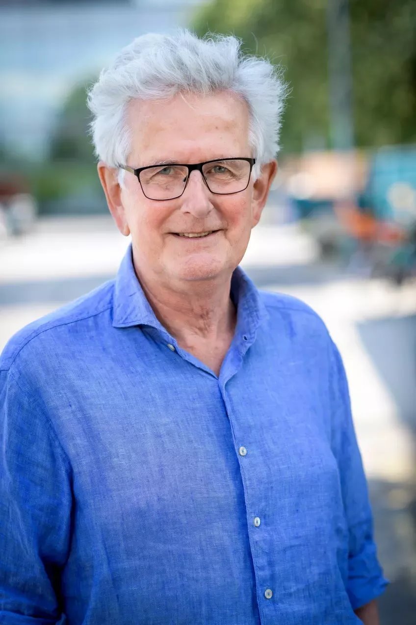
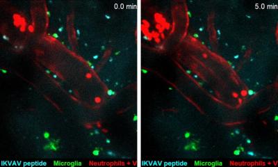
Image source: Lund University
“Communication between nerve cells in large parts of the brain changes after a stroke and we show that it can be partially restored with the treatment”, says Tadeusz Wieloch, senior professor of neurobiology at Lund University in Sweden.
In an ischemic stroke, lack of blood flow to the brain causes damage, which rapidly leads to nerve cell loss that affects large parts of the the vast network of nerve cells in the brain. This may lead to loss of function such as paralysis, sensorimotor impairment and vision and speech difficulties, but also to pain and depression. There are currently no approved drugs that improve or restore the functions after a stroke, apart from clot-dissolving treatment in the acute phase (within 4.5 hours of the stroke). Some spontaneous improvements occur, but many stroke patients suffer chronic loss of function. For example, about 60% of stroke sufferers, experience lost somatosensori functions such as touch and position sense.
An international study published recently in the journal Brain and led by a research team from Lund University in collaboration with University of Rome La Sapeinza and Washington University at St. Louis, shows promising results in mice and rats that were treated with a class of substances that inhibit the metabotropic glutamate receptor (mGluR5), a receptor that regulates communication in the brain’s nerve cell network.
The receptor mGluR5 is apparently an important factor in the reduced activity in the connectome, which is prevented by the inhibitor which therefore restores the lost brain function
Tadeusz Wieloch
“Rodents treated with the GluR5 inhibitor regained their somatosensori functions,” says Tadeusz Wieloch, who led the study.
Two days after the stroke, i.e. when the damage had developed and function impairment was most prominent, the researchers started treating the rodents that exhibited the greatest impaired function. “A temporary treatment effect was seen after just 30 minutes, but treatment for several weeks is needed to achieve a permanent recovery effect. Some function improvement was observed even when the treatment started 10 days after a stroke,” says Tadeusz Wieloch.
Importantly, sensorimotor functions improved, even though the extent of the brain damage was not diminished. This, explains Tadeusz Wieloch, is due to the intricate network of nerve cells in the brain, known as the connectome, i.e. how various areas of the brain are connected and communicate with each to form the basis for various brain functions. “Impaired function after a stroke is due to cell loss, but also because of reduced activity in large parts of the connectome in the undamaged brain. The receptor mGluR5 is apparently an important factor in the reduced activity in the connectome, which is prevented by the inhibitor which therefore restores the lost brain function,” says Tadeusz Wieloch.
The results also showed that sensorimotor function was further improved if treatment with the mGluR5 inhibitor is combined with somatosensory training by housing several rodents in cages enriched with toys, chains, grids, and plastic tubes.
The researchers hope that in the future their results could lead to a clinical treatment that could be initiated a few days after an ischemic stroke. “Combined with rehabilitation training, it could eventually be a new promising treatment. However, more studies are needed. The study was conducted on mice and rats, and of course needs to be repeated in humans. This should be possible since several mGluR5 inhibitors have been studied in humans for the treatment of neurological diseases other than stroke, and shown to be tolerated by humans,” says Tadeusz Wieloch.
Source: Lund University
05.12.2023