Article • On a shoestring budget
Bridging the resource gap for pathology in developing countries
Delivering a pathology service in resource-constrained locations and developing countries remains a challenge. Cost is a significant barrier, as is the availability of equipment, trained staff and technical and IT support can also hinder a desire from clinicians and pathologists to give their patients a high level of service to help their diagnosis and recovery. The subject was tackled in a specific session at the 37th European Congress of Pathology in September with speakers addressing the challenges and looking at steps to overcome them.
By Mark Nicholls
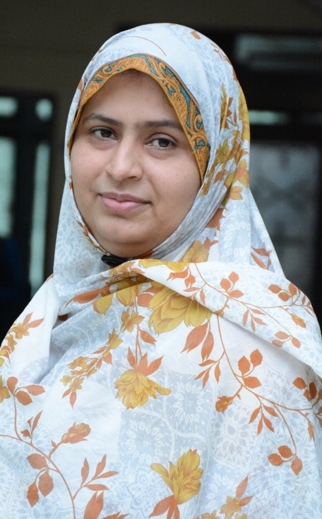
Image source: Jinnah Sindh Medical University
Two presentations at the congress highlighted different aspects of this challenge. Talat Zehra discussed how AI can contribute to genitourinary (GU) pathology in a resource-poor setting with her account of the challenges and potential solutions within Pakistan while Maysa Al-Hussaini from Jordan assessed the availability of advanced diagnostic methods in developing countries and presented findings from the Asian Oceanic Society of Neuropathology Survey.
Adapting digital pathology to local realities
Acknowledging how digitisation has revolutionised histopathology, with a need for scanners, digital microscopes, high resolution monitors, robust software, image management systems, strong IT infrastructure and cloud service, Dr Zehra reflected on how such expensive equipment is not readily available in lower resource settings. However, she added: ‘There are some solutions; while they are not the substitute for a scanner, these tools can help the pathologist to start their digital pathology journey.’
Zehra, who is Associate Professor at Liaquat National Hospital Medical College in Karachi, illustrated how a microscope equipped with a camera and connected to a screen enabled clinicians to view and share a digital image, even on mobile phone. She detailed a project in which her facility used an AI-based deep learning model for detecting metastatic deposits in lymph nodes of colorectal cancer with non-WSI images. ‘The aim was to compare the results obtained by the deep learning model with those determined by pathologists, assessing the agreement between the two methods,’ added Zehra.
Validating the low-cost approach
From 165 images, positive tumour areas were identified and despite the study’s small sample size, the results were promising. In addition, her team used an open-source software for stitching the single core biopsies of prostate and kidney and used it for research, education and knowledge sharing. They made videos at 10x through the camera-connected microscope and uploaded into open-source software and created special stains including trichome, PAS, silver stains and renal biopsy at 10x and 20x.
Advantages, she said, were that the whole slide images can be shared through links accessible on any screen including smart phones and can be shared for education, research and even telepathology. But there were limitations too – only single core biopsies can be used, there was an issue of blurring after 20x, and a file size of more than 3GB cannot be uploaded.
From bladder parasites to prostate cancer
The journey is continuous and we are hopeful
Talat Zehra
Her team also successfully used AI identification of schistosomiasis eggs in urinary bladder tissue with YOLOv8 variants, setting new performance benchmarks for the task. She said: ‘This technology offers a scalable and reliable diagnostic tool for resource-limited areas.’
Limited data availability and computational complexity of larger models can be a barrier for deployment on low-resource devices but future plans include expanding the dataset to include more diverse egg morphologies and imaging conditions and explore model optimisations for deployment on portable, low power, devices.
They also used AI in identification of perineural invasion of prostatic adenocarcinoma, with a high level (96.7%) of precision. ‘This was our small effort towards digitisation and use of AI tools in available resources,’ she added. ‘The journey is continuous and we are hopeful.’
Brain tumour diagnostic challenges
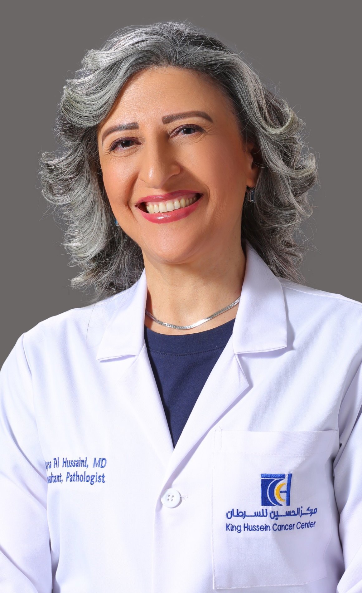
Image source: King Hussein Cancer Foundation and Cancer Center
Dr Maysa Al-Hussaini, Acting Chair of the Department of Cell Therapy and Applied Genomics, and a Neuropathologist at the King Hussein Cancer Center in Jordan, looked at the epidemiology of Central Nervous System (CNS) tumours, the most common tumour related cause of death in young people, and the challenges faced in low-resource settings, particularly in Asia and Africa. She shared findings from the Asian Oceanic Society of Neuropathology (AOSNP) Survey, looking at availability of molecular diagnostics in low, low-middle, high-middle and high-income countries within the Oceanian Region.
The AOSNP-ADAPTR (Adapting Diagnostic Approaches for Practical Taxonomy in Resource-Restrained Regions)1 found challenges to WHO CNS5, the World Health Organization’s 2021 classification for CNS tumours that integrated molecular diagnostics with traditional histological and immunohistochemical findings, in lower resource settings. These included lack of infrastructure, consumables, skilled personnel, validation, access to treatment, and cost.
Missing: Infrastructure, funding, training
In a study conducted by African and Asian neurosurgeons, 110 responses received from 92 countries within the Asian African region, showed that molecular diagnostics was not available. ‘Neuropathology and financial support stood as the major hinders for assigning or having a complete brain tumour programme,’ she said. ‘Twinning and training were proposed as possible solutions in order to overcome the shortage of molecular diagnostics.’
One third of African countries and institutions did not even have basic components for a neurosurgical programme. Further research underlined the lack of finance, molecular diagnostics and insufficient training.
The survey conducted by the AOSNP received 318 responses from institutions across 19 countries within the Asia-Oceania region and showed that basic and advanced molecular diagnostics was not available.
She said the AOSNP was establishing guidelines to support neuropathologists and neurosurgeons in such regions. ‘What we hope for is to have histology-oriented integrated diagnosis rather than for the integrated diagnosis to be only based on molecular diagnostics and some basic immunohistochemistry in order to help the pathologist and treating physician to reach the correct diagnosis,’ Al-Hussaini explained. ‘But there is a gap in the availability of diagnostic and treatment resources globally.’
The AOSNP-ADAPTR recommendations represent a response to an urgent need to address levels of resourcing and aim to ‘narrow the gap between better and less resourced countries.’
Profiles:
Dr Talat Zehra is Associate Professor at Liaquat National Hospital Medical College in Karachi, Pakistan.
Dr Maysa Al-Hussaini is Full Member, Neuropathologist and Acting Chair of the Department of Cell Therapy and Applied Genomics at the King Hussein Cancer Center in Amman, Jordan.
24.11.2025