Image source: Beizaee F, Lodygensky GA, Adamson CL et al., Medical Image Analysis 2025 (CC BY-NC 4.0)
News • Imaging consistency and reliability
Harmonizing MRIs across different institutions
Researchers at the Université de Montréal-affiliated Saint-Justine Hospital and the École de technologie supérieure (ETS) have come up with a better way to compare magnetic resonance images taken at different institutions.
Magnetic resonance imaging (MRI) is an essential tool for medical clinicians, providing detailed views of the interior of the human body as well as valuable information on pathologies. However, the variability of image acquisition protocols between different institutions poses significant challenges to achieving consistent and reliable interpretation, particularly in multi-centre research.
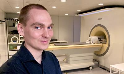
To solve this problem, a new study has been carried by Dr. Gregory Lodygensky, a clinical professor at Université de Montreal and clinician-researcher at its affiliated Sainte-Justine Hospital, with professor-researchers Jose Dolz and Christian Desrosiers of the École de technologie supérieure (ETS). Published in Medical Image Analysis, their study proposes modifying MRIs from different hospitals to make them more similar, enabling more reliable and accurate comparisons.
The analysis of these large cohorts in children and adults was hampered by the major harmonization problem, which has now been resolved
Gregory Lodygensky
Harmonization of MRI results is a central issue for research and health-care quality. Each hospital, clinic or research institute has its own particular MRI style, depending on the equipment, imaging protocols and parameters they use. This leads to variability in contrast, brightness and other image characteristics, and poses a major obstacle in clinical research when data from several research centres are pooled.
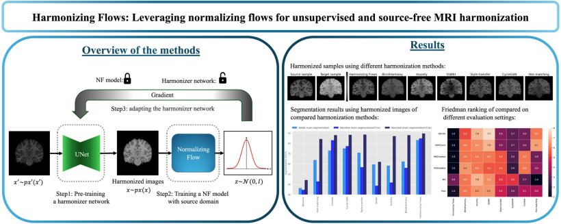
Developed by Farzad Beizaee, the study's first author and an ETS doctoral candidate, the new harmonization method involves three key steps:
- First, a model is created that “learns” how images in the source domain (for example, MRI images from a particular machine at Sainte-Justine) are organized or distributed.
- Once the distribution of the source domain is well understood, the aim is to “re-format” MRIs from other centres to eliminate variations caused by changes in parameters or the use of another machine, while at the same time preserving inherent patient differences.
- Lastly, when the model is used on new images (for example, from an unfamiliar machine), it must adapt and ensure that the new images still respect the distribution it learned in the first stage.
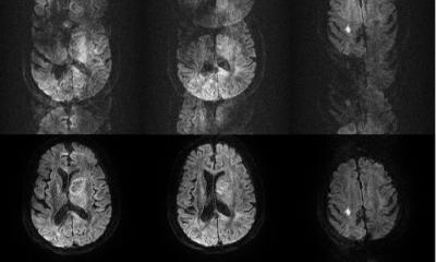
To validate their model, the researchers tested the new approach on MRI brain images held in databases in the United States and from a neonatal imaging consortium built in collaboration with researchers in Australia. These data were used to perform two different tasks: firstly, to segment brain images into different parts in adults and newborns to check whether brain structure remained consistent before and after harmonization, and secondly, to estimate brain age in newborns.
Recommended article

Article • Focus on radiology
Magnetic resonance imaging (MRI)
Imaging without ionising radiation: MRI uses magnetic fields to look inside the body. Keep up-to-date with the latest research news, medical applications, and background information on MR imaging.
The results highlighted the superior performance of this technique compared with existing harmonization methods, demonstrating its adaptability for a variety of tasks and population groups. Notably, the tool was successfully validated on the MRI of a newborn’s brain that had lesions, a task that all other available models fail to do since they are trained on images of healthy brains. “Thanks to this model, we can now interpret data from several thousands of families and children who are monitored at various hospitals – data that come from different scanners," said Lodygensky. "The analysis of these large cohorts in children and adults was hampered by the major harmonization problem, which has now been resolved.”
In future collaborations and research, he and his team will explore applying this approach on a larger scale, facilitating the comparison and analysis of research data and further improving the accuracy and reliability of medical diagnoses.
Source: Université de Montreal
03.03.2025