Image sources: left: Slivnick JA, Hawkes W, Oliveira J et al., European Heart Journal 2025 (CC BY 4.0); center: Feuchtner GM, Lacaita PG, Bax JJ et al.; Circulation: Cardiovascular Imaging 2025 (CC BY 4.0); right: Bhuva AN, Bai W, Lau C et al.; Circulation: Cardiovascular Imaging 2019 (CC BY-NC-ND 4.0)
Article • Expert perspectives from ESC 2025
Faster, smarter, deeper: how new technologies redefine cardiac imaging
Cardiac imaging is evolving, and new techniques continue to uncover the secrets of the heart for cardiologists who know how to use them. At the ESC 2025 Congress in Madrid, four experts explored cutting-edge developments across different modalities. Ranging from AI-assisted ultrasound image acquisition and accelerated MRI protocols to advanced prognostic tools for CT and nuclear imaging, these novel approaches provide a promising glimpse of what the next generation of cardiac imaging is capable of.
Article: Wolfgang Behrends
Starting off the expert panel, William A. Zoghbi, MD, pointed out how artificial intelligence can help flatten the notoriously steep learning curve of ultrasound image acquisition. This technology, which the expert from the Houston Methodist DeBakey Heart & Vascular Center, USA, likened to “image quality metre, almost like Wi-Fi bars”, provides automated guidance on how to obtain the relevant images for rheumatic heart disease screening.1 ‘This will have quite a bit of impact for novices, increasing the time for expertise and the availability in rural areas and underserved areas,’ he predicted, ‘not only for rheumatic heart disease, but also in heart failure and any other screening.’
AI is not a threat, but it is indeed an opportunity for echocardiography to grow and have more impact
William A. Zoghbi
Other advances also bring benefits to more experienced sonographers, such as AI-based quantification of cardiac volumes and automated reporting.2 Even though there will still be the need for a human to verify the AI-generated content, this will significantly reduce examination times, Dr Zoghbi emphasized. Building on this foundation, algorithms have even taken on disease detection and interpretation, he continued, highlighting a recent model capable of detecting cardiac amyloidosis from a single video.3
Recommended article
News • Neural network screening tool
AI-enhanced ultrasound early detects cardiac amyloidosis
A neural network AI has been trained to detect cardiac amyloidosis from a single echocardiogram video of the heart's apical four-chamber view and differentiate it from similar heart conditions.
Moving from detection to prediction, cardiologists should harness the technology’s ability to extrapolate data from multiple sources for risk assessment, the expert advised: ‘That’s what patients really care about.’4
So, will echocardiography thrive with the new technology or suffer from its impact? For Dr Zoghbi, the answer is clear: ‘AI is not a threat, but it is indeed an opportunity for echocardiography to grow and have more impact,’ he concluded. ‘It is really amazing that every few years, there is a new innovation in echocardiography that not only allows us to see and understand the heart better but hopefully also help take care of patients and improve their overall outcome.’
Cardiac CT: less radiation, fewer artifacts, more applications
This enthusiasm was shared by Prof. Dr Dr Christoph Gräni. Speaking as a champion for CT, he had no difficulty in listing the many advances the modality – which already had a strong standing in cardiac imaging to begin with – underwent in recent years: Adding to its high acquisition speed, new technologies greatly improved image quality while reducing radiation exposure to a minimum. The Head of Cardiac Imaging at Bern University Hospital, Switzerland, was particularly impressed with the prognostic value: ‘From a normal coronary CT, we can give the patient a guarantee of ten years that he or she doesn't have a myocardial infarction – that's a very strong statement.’5
On the hardware side, the introduction of photon-counting CT has significantly pushed the diagnostic capabilities. The new technology increases resolution, but also reduces imaging artifacts like blooming, Gräni observed, allowing for new insight in patients with coronary calcifications or stents.6
Recommended article
News • Classification for coronary artery disease
Study: Photon-counting CT improves CAD assessment
Higher image quality, better spatial resolution: A new radiology study demonstrates how photon-counting CT imaging can improve evaluation of coronary artery disease.
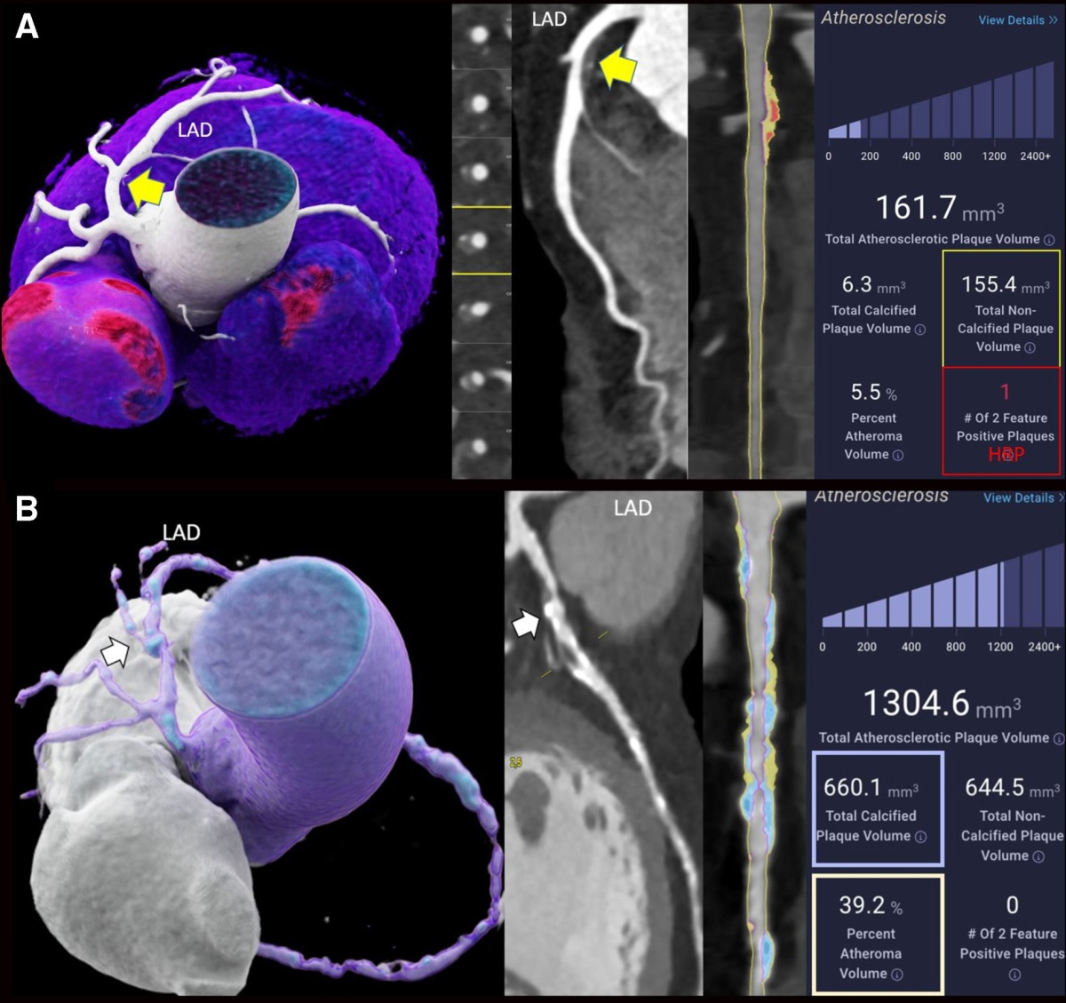
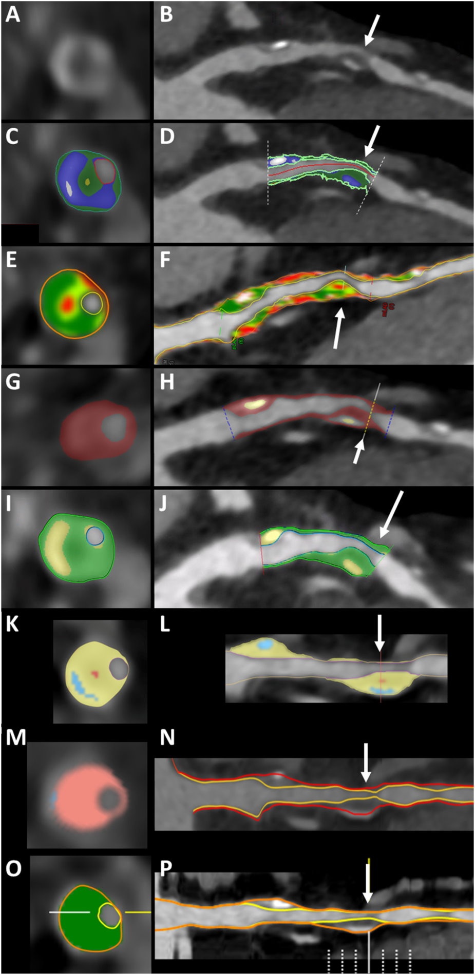
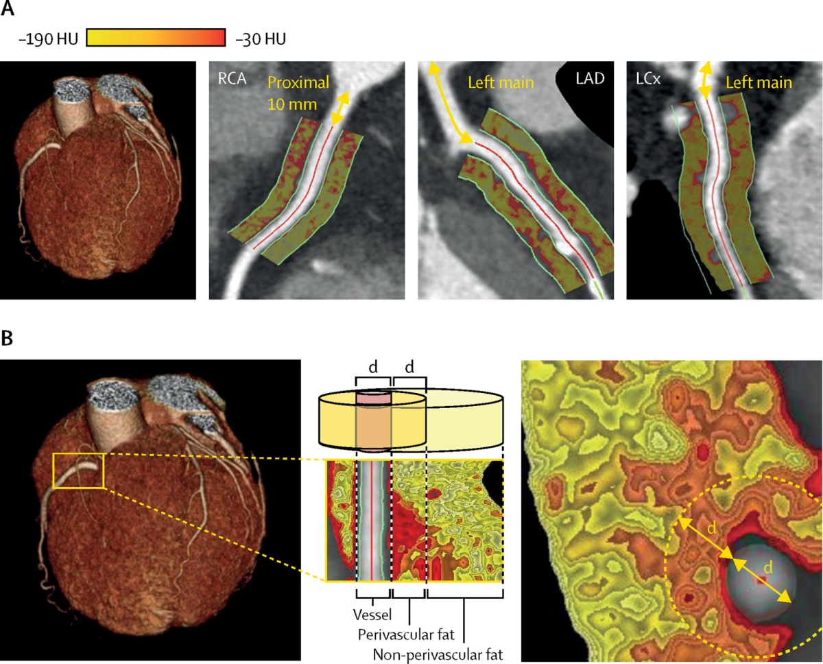
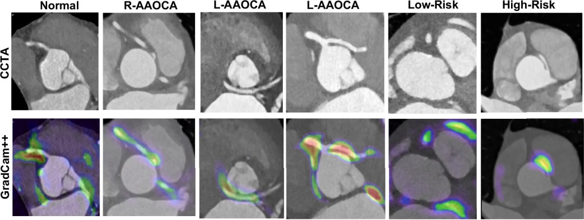
‘It's mainly the scanner technology which has evolved but also the post-processing,’ he said. ‘And this entire journey is accelerated and improved by artificial intelligence.’ Among the many promising CT applications for AI, the expert highlighted solutions for reducing inter-reader variability7, harnessing the prognostic potential of coronary plaque and fat attenuation8,9,10, and detecting rare diseases like coronary artery anomalies.11
Another exciting development the expert pointed out is in simulating procedure outcomes: Whereas prior approaches already implemented data-driven digital twins, more recent research has turned to producing 3D-printed physical twins for hemodynamic stress testing.12 ‘You do a 3D printing out of the CT scan and then you apply it into a flow loop,’ Gräni explained. In the future, he predicted, these physical twins will help cardiologists to predict coronary obstructions, ruptures, help with lifetime management of cardiac implants, and much more. How far into the future? The expert is convinced that ‘these patient-specific digital and physical twins, I'm sure will come very soon’.
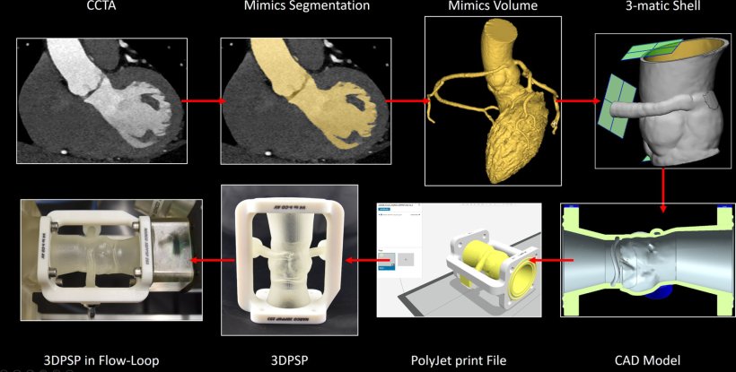
Image source: Illi J, Stark AW, Ilic M et al.; JACC: Case Reports 2024 (CC BY 4.0)
CMR: advances in speed and user-friendliness
With such strong competition, will cardiac MRI (CMR) be left behind? According to Dr Chiara Bucciarelli-Ducci, this modality is neither obsolete – nor is it slow: ‘CMR can be quick, and it is quick if you know what you're doing,13’ the cardiologist from Royal Brompton and Harefield Hospital, UK, stated. And not only is it fast, but it also delivers clinically relevant information, she pointed out. Using the right protocols, comprehensive stress CMR for ischemic heart disease investigation can reliably be performed in well under 20 minutes.14 Add new features such as one-click image assessments15 and AI-assisted auto-contouring16, and cardiologists have a very powerful modality at their hands: ‘When I started CMR 20 years ago, it was taking an hour to analyse and was very operator-dependent,’ the expert said. ‘Now, you do one click and all the AI does the contouring for you, not just of the volumes but also the late enhancement.’
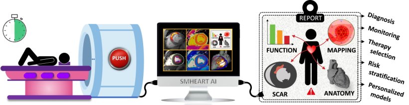
Image source: Bustin A, Stuber M, Sermesant M, Cochet H; European Heart Journal 2023 (CC BY-NC 4.0)
The future of the modality is bright as well, according to the Bucciarelli-Ducci: From CMR fingerprinting for simultaneous and reproducible measurement of T1 and T2 maps in a single scan17, multitasking for setup-free imaging18, quantitative perfusion imaging to non-invasively diagnose ischaemia19, to oxygenation-sensitive CMR for contrast- and needle-free heart failure diagnostics20 – there are plenty of new approaches to improve the already strong foundation and close the implementation gap. ‘By making CMR quicker, easier, empowering more users, and making it safer, we can work towards making it as available and as fast as CT – and we’re getting there,’ she concluded.
Nuclear imaging: seeing the cardiovascular system as a whole
Compared to MRI, which is considered the first-line diagnostic tool for a wide range of cardiovascular conditions, nuclear imaging plays a less prominent role. Despite its niche role, however, the modality should not be overlooked, said Marc Dweck, Professor of Clinical Cardiology and Consultant at the University of Edinburgh and final speaker of the expert panel. ‘Actually, I think nuclear imaging has the most exciting future compared to other techniques, at least in terms of the incremental information that I think it's going to provide us over the next few years,’ he shared.
[Having these new tracers] opens up really interesting opportunities to better understand disease, but also to make strides about improving diagnosis, assessing response to therapy, and more
Marc Dweck
Molecular PET imaging, with its ability to not only assess anatomy of cardiac function, but monitor the activity of specific processes, is particularly promising in this regard. For example, FDG PET imaging is already used to detect inflammation and infection in prosthetic valve endocarditis and cardiac sarcoidosis.21 New tracers like 18F-fluoride, 18F-fluoride GP1, and FAPI expand on this by providing even more detailed insights into inflammation, thrombus formation, calcification activity, and fibrosis activity.22 ‘Now we have tracers that inform about each of the major pathologies or pathological processes that drive cardiovascular disease,’ Dweck pointed out. ‘This opens up really interesting opportunities to better understand disease, but also to make strides about improving diagnosis, assessing response to therapy, and more.’ This is especially relevant for diseases with no clear underlying cause, such as stroke, he added.
The advent of total body PET scanners is another milestone for nuclear imaging, enabling a comprehensive look at the cardiovascular system as a whole. ‘We can look at the brain, we can look at the heart, we can look at the peripheral vasculature,’ Dweck said. ‘And it allows us to look at interactions between different organ systems. If we combine that with the amazing new tracers that we have, I hope you will now agree that this is the most exciting cardiovascular imaging technology for the future.’
Who does the future of cardiac imaging belong to?
So, how do these advances translate to future cardiac imaging? Maybe Gräni put it best when he concluded: ‘Multimodal imaging is key; that's what we do nowadays, but that will also be the future. We have the patient in the centre with a clinical question and we try to best answer that by deciding which modality or which combination of modalities we choose. So, there's no one best modality or one modality wins over the other.’
Profiles:
William A. Zoghbi, MD, is the Elkins Family Distinguished Chair in Cardiac Health, chair of the Department of Cardiology at Houston Methodist DeBakey Heart and Vascular Center, and Professor of Medicine at Weill Cornell Medical College and the Houston Methodist Academic Institute, USA. He has dedicated his career to advancements in clinical cardiology, enhancing non-invasive approaches to assess and managing cardiac disease through cardiovascular imaging. His areas of expertise include valvular heart disease, coronary artery disease and heart failure, emphasizing a patient-centered approach to medicine and general cardiology.
Dr Christoph Gräni is Professor in Cardiology at the University of Bern, Switzerland, and Director of Cardiac Imaging, covering Echocardiography, Cardiac Magnetic Resonance, Cardiac Computed Tomography and Nuclear Cardiology at the University Hospital Bern. One of his main research focuses is on improving the diagnosis and risk stratification of different cardiomyopathies, myocarditis and cardiac amyloidosis, using multimodality cardiac imaging myocardial function analysis and fibrosis assessment. Additionally, his research includes non-invasive assessment of coronary artery disease, especially coronary artery anomalies.
Dr Chiara Bucciarelli-Ducci is a cardiologist from Royal Brompton and Harefield Hospital, UK and associate professor at King College London’s School of Biomedical Engineering & Imaging. She is an international opinion leader on the use of CMR in cardiovascular medicine and has over 20 years of clinical experience and expertise. Her career focuses on improving the care of patients by improving the identification of heart abnormalities in a wide range of conditions and symptoms.
Dr Marc Dweck is Professor of Clinical Cardiology and Consultant at the University of Edinburgh, UK. He is Vice President of EACVI with clinical interests in multi-modality imaging and cardiac device implantation. His research program is centred around the use of multi-modality imaging (echo, CT, CMR, PET) to improve our understanding of cardiovascular pathophysiology and ultimately to improve patient assessment, care and outcomes. He has published in many of the leading medical and cardiovascular journals and is the recipient of numerous national and international awards.
References:
- Peck D, Rwebembera J, Nakagaayu D et al.: The Use of Artificial Intelligence Guidance for Rheumatic Heart Disease Screening by Novices; Journal of the American Society of Echocardiography 2023; https://doi.org/10.1016/j.echo.2023.03.001
- Hirata Y, Nomura Y, Saijo Y, Sata M, Kusunose K: Reducing echocardiographic examination time through routine use of fully automated software: a comparative study of measurement and report creation time; Journal of Echocardiography 2024; https://doi.org/10.1007/s12574-023-00636-6
- Slivnick JA, Hawkes W, oliveira J et al.: Cardiac amyloidosis detection from a single echocardiographic video clip: a novel artificial intelligence-based screening tool; European Heart Journal 2025; https://doi.org/10.1093/eurheartj/ehaf387
- Valsaraj A, Kalmady SV, Sharma V et al.: Development and validation of echocardiography-based machine-learning models to predict mortality; eBioMedicine 2023; https://doi.org/10.1016/j.ebiom.2023.104479
- Finck T, Hardenberg J, Will A et al.: 10-Year Follow-Up After Coronary Computed Tomography Angiography in Patients With Suspected Coronary Artery Disease; JACC Cardiovascular Imaging 2018; https://doi.org/10.1016/j.jcmg.2018.07.020
- Sakai K, Shin D, Singh M et al.: Diagnostic Performance and Clinical Impact of Photon-Counting Detector Computed Tomography in Coronary Artery Disease; JACC 2024; https://doi.org/10.1016/j.jacc.2024.10.069
- Feuchtner GM, Lacaita PG, Bax JJ et al.: AI-Quantitative CT Coronary Plaque Features Associate With a Higher Relative Risk in Women: CONFIRM2 Registry; Circulation: Cardiovascular Imaging 2025; https://doi.org/10.1161/CIRCIMAGING.125.018235
- Nieman K, García-García HM, Hideo-Kajita A et al.: Standards for quantitative assessments by coronary computed tomography angiography (CCTA); Journal of Cardiovascular Computed Tomography 2024; https://doi.org/10.1016/j.jcct.2024.05.232
- Oikonomou EK, Marwan M, Desai MY et al.: Non-invasive detection of coronary inflammation using computed tomography and prediction of residual cardiovascular risk (the CRISP CT study): a post-hoc analysis of prospective outcome data; Lancet 2018; https://doi.org/10.1016/S0140-6736(18)31114-0
- Chan K, Wahome E, Tsiachristas A et al.: Inflammatory risk and cardiovascular events in patients without obstructive coronary artery disease: the ORFAN multicentre, longitudinal cohort study; Lancet 2024; https://doi.org/10.1016/S0140-6736(24)00596-8
- Shiri I, Baj G, Mohammadi Kazaj P et al.: AI-based detection and classification of anomalous aortic origin of coronary arteries using coronary CT angiography images; Nature Communications 2025; https://doi.org/10.1038/s41467-025-58362-9
- Illi J, Stark AW, Ilic M et al.: Hemodynamic Relevance Evaluation of Coronary Artery Anomaly During Stress Using FFR/IVUS in an Artificial Twin; JACC: Case Reports 2024; https://doi.org/10.1016/j.jaccas.2024.102729
- Raman SV, Markl M, Patel AR et al.: 30-minute CMR for common clinical indications: a Society for Cardiovascular Magnetic Resonance white paper; Journal of Cardiovascular Magnetic Resonance 2022; https://doi.org/10.1186/s12968-022-00844-6
- Foley JRJ, Richmond C, Fent GJ et al.: Rapid Cardiovascular Magnetic Resonance for Ischemic Heart Disease Investigation (RAPID-IHD); JACC: Cardiovascular Imaging 2020; https://doi.org/10.1016/j.jcmg.2020.01.029
- Bustin A, Stuber M, Sermesant M, Cochet H: Smart cardiac magnetic resonance delivering one-click and comprehensive assessment of cardiovascular disease; European Heart Journal 2023; https://doi.org/10.1093/eurheartj/ehac814
- Bhuva AN, Bai W, Lau C et al.: A Multicenter, Scan-Rescan, Human and Machine Learning CMR Study to Test Generalizability and Precision in Imaging Biomarker Analysis; Circulation: Cardiovascular Imaging 2019; https://doi.org/10.1161/circimaging.119.009214
- Liu Y, Hamilton J, Rajagopalan S et al.: Cardiac Magnetic Resonance Fingerprinting: Technical Overview and Initial Results; JACC: Cardiovascular Imaging 2018; https://doi.org/10.1016/j.jcmg.2018.08.028
- Christidoulou AG, Shaw JL, Ngyuen C et al.: Magnetic resonance multitasking for motion-resolved quantitative cardiovascular imaging; Nature biomedical Engineering 2018; https://doi.org/10.1038/s41551-018-0217-y
- Kotecha T, Martinez-Naharro A, Boldrini M et al.: Automated Pixel-Wise Quantitative Myocardial Perfusion Mapping by CMR to Detect Obstructive Coronary Artery Disease and Coronary Microvascular Dysfunction: Validation Against Invasive Coronary Physiology; JACC: Cardiovascular Imaging 2019; https://doi.org/10.1016/j.jcmg.2018.12.022
- Hillier E, Friedrich MG: The Potential of Oxygenation-Sensitive CMR in Heart Failure; Current Heart Failure Reports 2021; https://doi.org/10.1007/s11897-021-00525-y
- Pizzi MN, Roque A, Cuéllar-Calabria H et al.: 18F-FDG-PET/CTA of Prosthetic Cardiac Valves and Valve-Tube Grafts: Infective Versus Inflammatory Patterns; JACC: Cardiovascular Imaging 2016; https://doi.org/10.1016/j.jcmg.2016.05.013
- Rubeaux M, Joshi NV, Dweck MR et al.: Motion Correction of 18F-NaF PET for Imaging Coronary Atherosclerotic Plaques; Journal of Nuclear Medicine 2016; https://doi.org/10.2967/jnumed.115.162990
13.10.2025