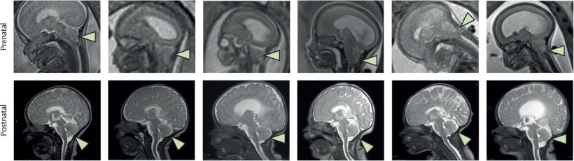
Image source: Farmer DL, Kumar P, Reynolds E et al., The Lancet 2026 (CC BY-NC-ND 4.0)
News • Combining stem cell therapy with surgery
Spina bifida: in-utero stem cell treatment approach shows promise
New approach could lead to better health outcomes for babies compared to traditional fetal surgery
This is the first time live stem cells have been used on a fetus’ damaged spine, which could potentially lead to better health outcomes for babies compared to traditional fetal surgery.
Results of the phase 1 clinical trial were published in The Lancet.
Spina bifida is a congenital condition in which the spinal cord does not develop properly, leaving part of it exposed. This can cause lifelong health challenges, including paralysis, difficulty walking, and issues with bladder and bowel control. Current treatments involve surgery during pregnancy to close the spinal opening, which can reduce some complications but often does not prevent all neurological problems.
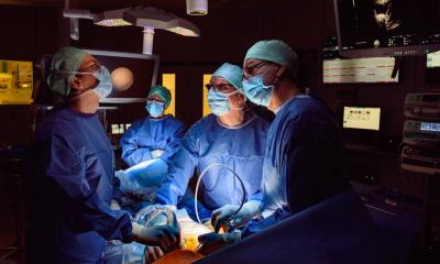
In the trial, six pregnant women with fetuses diagnosed with spina bifida underwent standard fetal surgery plus an additional step where surgeons applied cells from the placenta—called placenta-derived mesenchymal stem cells (PMSCs)—directly onto the exposed spinal cord during the surgery. These cells are known for their ability to reduce inflammation, promote healing, and protect nerve tissue.
All six babies, who were born between July 2021 and December 2022, had their spinal repairs intact and showed no signs of infection, abnormal tissue growth, or tumour formation. Post-birth MRI scans confirmed that brain abnormalities associated with spina bifida, called hindbrain herniation, were reversed in every case.
In addition, the infants experienced no serious adverse effects attributable to the stem cell treatment during the trial and through follow-up care. The children in this trial will be carefully monitored, with regular check-ups and assessments, until they turn six. This long-term follow-up will help researchers confirm that the stem cell treatment remains safe and improves children’s mobility, health, and quality of life as they grow.
Additional large-scale, long-term clinical trials are underway to further refine surgical techniques and treatment protocols. These studies aim to confirm that children who receive this therapy experience benefits at birth, improved mobility, and a better quality of life. Throughout this process, regulatory agencies will work closely with the research team to carefully monitor safety and effectiveness.
The researchers aim to establish this stem cell therapy as a safe and standard option for fetal repair of spina bifida, providing new hope for families affected by this condition worldwide. They say these results represent a major milestone in the field of in-utero stem cell treatment for birth defects, paving the way for future advances using stem cells during fetal surgery to address other congenital conditions.
Source: The Lancet
27.02.2026