Article • Affordable, sustainable – but underused
Low-field MRI: The imaging solution radiologists haven't learned to trust
At the 2025 ESMRMB Annual Meeting in Marseille, speakers made a strong case for what remains an outsider in radiology: low-field MRI. Despite its affordability, improved performance, and reduced environmental footprint, the technology continues to face scepticism – not from regulators or patients, but from radiologists themselves.
By Mélisande Rouger
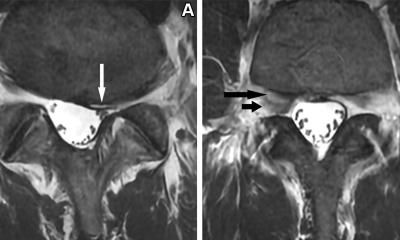
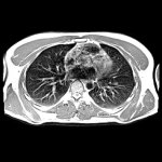
Image source: Pogarell T, Heiss R, Janka R, Nagel AM, Uder M, Roemer FW, Skeletal Radiology 2024 (CC BY 4.0)
‘Most residents are trained on 1.5T or 3T systems. They’ve never even seen a low-field scanner,’ said Dr Kai Vilanova, Director of Magnetic Resonance at Clínica Girona and Associate Professor at the University of Girona, Spain. ‘And so they grow up believing low-field equals low quality – which is simply not true.’

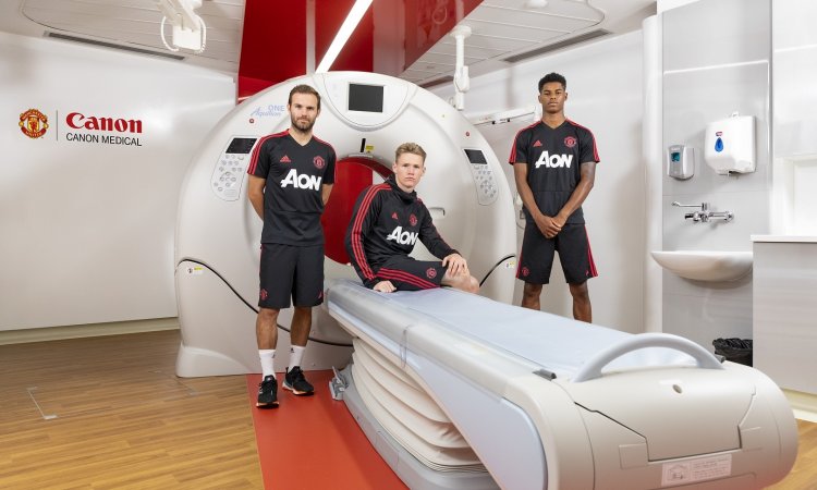
Image source: ESMRMB
Vilanova, who will chair the ESMRMB 2026 meeting, has made low-field implementation a cornerstone of his clinical strategy. In Marseille, he laid out a detailed case for its integration into routine practice: economic sustainability, sufficient diagnostic quality for MSK and spine imaging, and growing support from AI technologies.
‘In my clinic, nearly 80% of MRI requests are for spine or MSK,’ he explained. ‘The question is not whether we can produce beautiful images – the question is whether we can answer the clinical question.’
For knee pain, lumbar discomfort or degenerative joint changes, low-field MRI offers enough diagnostic clarity at a fraction of the cost of high-field scans. ‘We’re not talking about advanced cardiac or prostate imaging,’ he added. ‘We’re talking about the everyday core of radiology, where efficiency matters most.’
Vilanova’s facility in Girona runs a multimodal fleet: 3T, 1.5T, and low-field units, including upright systems. ‘We use 3T where it’s needed – but not for everything,’ he said. ‘For most cases, a compact car does the job. We don’t need a Ferrari just to drive around the block.’
Economic and environmental logic
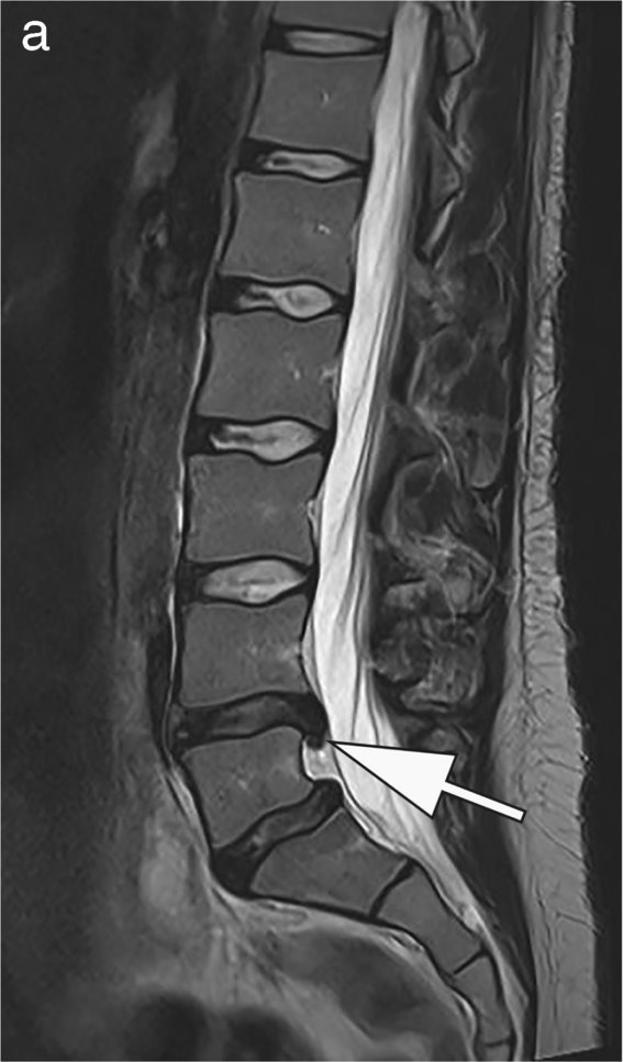
Image source: Pogarell T, Heiss R, Janka R, Nagel AM, Uder M, Roemer FW, Skeletal Radiology 2024 (CC BY 4.0)
Beyond clinical performance, low-field MRI offers compelling advantages in cost and sustainability. ‘Acquisition, maintenance, energy consumption – everything is four to five times cheaper than high-field,’ Vilanova noted. ‘And the diagnostic reimbursement is exactly the same.’
He pointed to a 2023 internal study comparing the cost-efficiency of different field strengths in his department. ‘Low-field emerged as the most sustainable choice – economically and ecologically,’ he said. ‘Especially for MSK and spine imaging, where high-resolution is often unnecessary.’
In Spain, around 200 low- and mid-field MRI systems are currently installed, accounting for approximately 15% of the national MRI fleet. Most are found in outpatient centres and smaller diagnostic units. ‘We’ve seen real growth in the last decade,’ he said. ‘But institutional resistance remains strong.’
According to Vilanova, the main obstacle isn’t technical – it’s cultural. ‘Public hospitals don’t install low-field machines. As a result, residents are never exposed to them,’ he said. ‘And if they don’t learn how to interpret lower-resolution images, they won’t trust the results. That’s the crux of the issue.’
He believes radiology must update its training models. ‘We are training radiologists to drive Ferraris. But healthcare systems also need drivers who can operate smart, agile vehicles. That flexibility should be part of our educational DNA.’
AI for the future potential of low-field MRI
AI can compensate reduced SNR, spatial resolution and longer scan times, he believes. ‘Early studies show AI-enhanced low-field MRI achieves similar diagnostic quality as high field systems,’ he said. ‘Cross-field harmonization models reduce variability between field strengths. AI acts as a “virtual amplifier”, balancing hardware limitations with software intelligence.’
The future of sustainable MRI will rely on the integration of low-field systems and AI-based reconstruction, he added.
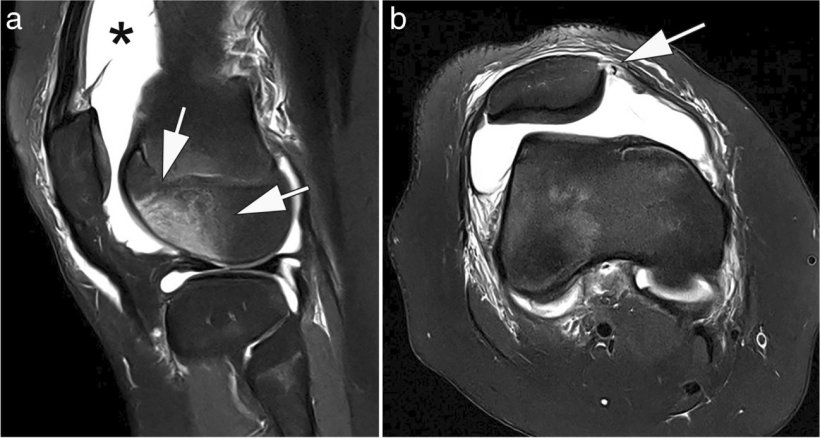
Image source: Pogarell T, Heiss R, Janka R, Nagel AM, Uder M, Roemer FW, Skeletal Radiology 2024 (CC BY 4.0)
A complement, not a replacement
Vilanova insists low-field MRI is not intended to replace high-end systems. ‘We still need 3T for advanced exams – cardiac, breast, functional neuro,’ he said. ‘But low-field should be used strategically, to offload high-field units and improve workflow.’
At ESMRMB 2026 in Girona, low-field applications will take center stage again – not just as a technology, but as a tool for rethinking resource allocation in radiology. ‘Low-field is clinically relevant, cost-effective, and sustainable,’ Vilanova concluded.
Pushing the limits even lower
If Vilanova champions low-field MRI, others are exploring what lies even further below. In Marseille, Dr Edmond Knopp, neuroradiologist and former Associate Professor at NYU Langone, New York, presented clinical experience with a portable ultra-low-field scanner (0.064T) dedicated to brain imaging.
Designed for bedside use in ICUs, operating theatres or emergency settings, the system challenges the traditional boundaries of MRI: ‘It’s mobile, it doesn’t need shielding, and it doesn’t require a specialized radiographer to operate,’ Knopp said. ‘We’re not just lowering field strength – we’re expanding where, how, and by whom MRI can be performed.’
Recommended article
Article • Expanding potential
Low-field MRI: Less is more
Currently, two opposing trends can be observed in MRI: on the one hand 1.5T scanners are increasingly replaced by 3T scanners for standard clinical MRI applications. On the other hand scanners with lower field strengths have become commercially available in the past years. A session at ECR 2021 took a closer look at low-field (0.5T) and ultra-low-field (0.05T) MRI scanners.
From stroke triage to post-operative follow-up, the scanner delivers actionable diagnostic data in settings where high-field MRI is logistically impossible. ‘Lowering the field doesn’t mean limiting diagnosis,’ Knopp concluded. ‘It means extending it to places – and patients – who didn’t have access before.’
Profiles:
Dr Kai Vilanova, MD, PhD, is Director of the Magnetic Resonance Institute at Clínica Girona, Consultant Radiologist at IDI–University Hospital of Girona, and Associate Professor at the University of Girona, Spain. He serves as Director of the ESMRMB School of MRI, and Chair of the ESMRMB Congresses (Barcelona 2024, Girona 2026). A former President of the Spanish Skeletal Radiological Society, he has authored over 150 papers and 9 books, with 250 invited lectures. His distinctions include the SERAM Research Award and the Career Achievement Award from the Medical Association of Girona.
Dr Edmond Knopp is an experienced neuroradiologist with more than 30 years of experience in both private and public institutions. He is also Co-Founder of Acumenhealth and Chief Medical Officer of Hyperfine. He served as Associate Professor of Radiology and Neurosurgery at NYU Langone Medical Center for over 20 years in New York, USA.
30.10.2025