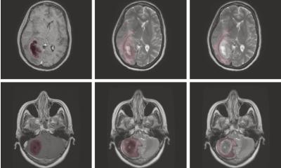
Image source: Mallikourti V et al., Radiology 2024 (CC BY 4.0)
News • Field-cycling imaging
FCI: New MRI-derived scanner shows promise for stroke imaging
A new type of medical scanner developed by a University of Aberdeen team has shown that it can identify brain damage in stroke patients at lower magnetic fields than ever before.
The world’s first Field Cycling Imager (FCI) derives from MRI but is able to work at ultra-low magnetic fields, which makes it capable of seeing how organs are affected by diseases in previously unseen ways. Because it is safer than current scanners, FCI also opens exciting possibilities for the development of systems tailored for ambulances and other out-of-hospital settings.
Magnetic resonance imaging (MRI) uses strong magnetic fields and radio waves to produce detailed images of the inside of the body without touching it. The FCI scanner is based on the same principles, but its design is completely revisited so that it can vary the strength of the magnetic field during the patient’s scan. This includes going down to magnetic fields of less than a fridge magnet, while obtaining good quality pictures. Being able to vary the magnetic field makes it like having multiple scanners in one and it can therefore extract more information - and different information - than traditional MRI.

Image source: University of Aberdeen
A paper published in Radiology shows that the stroke area due to a blocked blood vessel in the brain produces a consistently different signal from normal brain at very low magnetic field strengths – a factor of 10,000 lower than traditional MRI and 100 times lower that other low field systems.
Professor Mary Joan MacLeod, Professor of Stroke Medicine, said: “Our initial findings are very exciting, as they reflect the first step to producing a device that would be safe and small enough to put in an ambulance so that stroke patients can have a diagnosis and start treatment before they reach hospital. We have also shown that the new scanner can also identify bleeds in the brain, and changes in the small blood vessels which might lead to dementia. We know that there are lots of other exciting potential applications for this technology in areas such as cancer and bone disease. We would like to thank all the patients who have so enthusiastically helped us get to this stage”

Image source: University of Aberdeen
Dr Lionel Broche, senior Research Fellow in Biomedical Physics, said: “This success is the result of a long research effort and a strong collaboration between the University of Aberdeen and the stroke team at Aberdeen Royal Infirmary. As we keep improving the technology for field-cycling imaging, we are starting to see new signals with great potential for clinical applications. We are now planning the continuation of this study on stroke using the next design iteration of our field-cycling technology, which will be located directly inside the Aberdeen Royal Infirmary. This will make it directly accessible for more patients and will open many opportunities for medical research. These are exciting times.”
Professor David Lurie, Emeritus Professor of Medical Physics said: “It is fantastic that Field-Cycling Imaging, which the Aberdeen team has been developing for more than a decade, is now showing its value in the assessment of stroke. This work points the way forward for FCI, with benefits to patients and health services worldwide.”
Source: University of Aberdeen
16.12.2024


