News • Depth-resolved fiber photometry
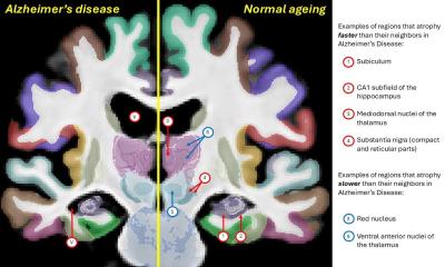
Tracking Alzheimer’s progression in real time
Scientists have developed an innovative technique to observe Alzheimer’s disease as it progresses in the brain – an important step forward that could accelerate new treatments.
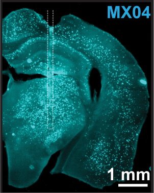
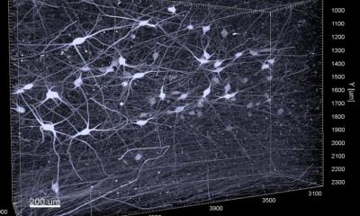
Image source: Byron N, McAlinden N, Pisano F et al., Neurophotonics 2025 (CC BY 4.0)
The new method uses light to detect and measure the build-up of amyloid plaques – a key characteristic of Alzheimer’s – at different depths in the brain. This is the first time such measurements have been made in living animals as they move naturally, rather than under anaesthetic or in fixed positions.
The research, published in the journal Neurophotonics, was carried out by the University of Strathclyde in partnership with the Istituto Italiano di Tecnologia, University of Padua, and the Technical University of Denmark, and combines expertise in neuroscience, physics and biomedical imaging.
A special fluorescent dye that attaches to amyloid plaques in the brain was used, with ultra-thin optical fibres then applied to shine light into the brain and measure how much of the dye was present – giving a clear picture of how much plaque had formed.
The system was tested in mice that had been genetically modified to develop Alzheimer’s-like symptoms. The results closely matched those seen in postmortem brain tissue, confirming that the new technique is accurate.
Professor Shuzo Sakata at Strathclyde and senior author of the study, said: "Being able to monitor changes in the brain as they happen – in real time and across different brain regions – is a major step forward. It will help researchers understand how Alzheimer’s disease develops and test whether new treatments are working, more quickly and accurately than before."
The research was funded by the European Union’s Horizon 2020, the Medical Research Council, part of UKRI, and Alzheimer’s Research UK.
Dr Niall McAlinden, co-author, and an expert in Photonics at Strathclyde, said: “This approach allows us to track the disease over time in the same subject, and opens new doors for studying the progression of Alzheimer’s and how it responds to treatment.”
The team is now working to improve the system’s capabilities and explore its use in other characteristics of Alzheimer’s disease.
Source: University of Strathclyde
09.10.2025