News • Algorithms put to the test
Diffusion MRI denoising: do sharper images equal better diagnostics?
Researchers from the National Institute of Physiological Sciences found that ‘denoising’, a method used to clean diffusion-weighted MRI (dMRI) data, alters the appearance of dMRI images and the estimated signal-to-noise ratio of the data, but had limited impacts on the ability to detect optic tract abnormalities in glaucoma.
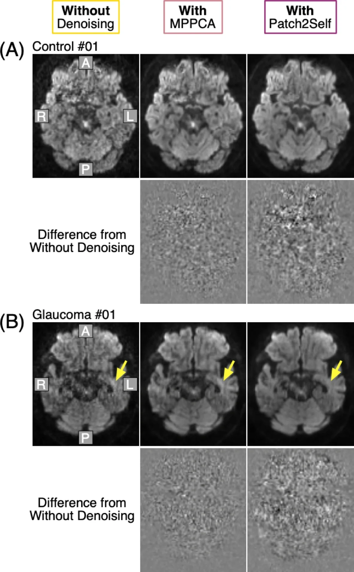
Image source: Takuma D, Ogawa S, Takemura H, Scientific Reports 2025 (CC BY-NC-ND 4.0)
The researchers published their insights in the journal Scientific Reports.
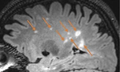
For decades, MRI (magnetic resonance imaging) has been a powerful way to see inside the human brain. A specialized form, diffusion-weighted MRI (dMRI), can measure tissue properties of fiber pathways in the brain by measuring the movement of water molecules. Since dMRI is applicable to living humans, it becomes a valuable tool for studying the neurological changes of fiber pathways caused by disorders.
However, dMRI data often suffers from the presence of ‘noise’, limiting its ability to identify biological signals related to health and disease. To date, several ‘denoising’ algorithms have been proposed to remove noise from dMRI data. Now, in a study to be published in Scientific Reports, researchers from the National Institute of Physiological Sciences have sought to determine in what aspect ‘denoising’ can help identify disease-related changes in fiber pathways. “We decided to check for ourselves if the available denoising techniques were truly beneficial for empirical data,” explains Daiki Taguma, lead author of the study.
The research team tested two common denoising techniques on real-world data: brain scans from healthy individuals and patients with glaucoma. Previous research had already shown that dMRI could detect tissue abnormalities in the fiber pathway named the optic tract in glaucoma, making it an ideal test case. They found that denoising altered the appearance of dMRI images and increased the estimated signal-to-noise ratio, a key metric for evaluating image quality. However, they also found that when they analyzed dMRI data of the optic tract, denoising had a limited impact on the differences between healthy controls and patients with glaucoma.
These results suggest that at present, denoising may improve dMRI data in some aspect, but does not significantly alter results concerning disease-related changes in fiber pathways. The study concluded that the benefits of current denoising methods depend on the context and purpose of the analysis.
These findings mark an important step toward refining how dMRI is used in both clinical and research settings, the researchers conclude. By understanding what works, researchers can better harness dMRI’s potential, advancing our ability to detect and understand the impact of diseases on fiber pathways in the brain.
Source: National Institute for Physiological Sciences
08.08.2025