News • Microscopy
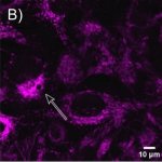
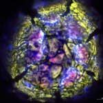
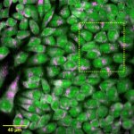
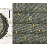
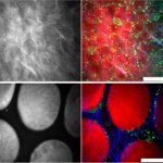
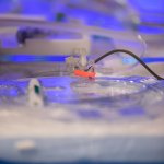
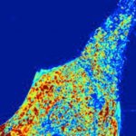
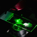
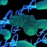
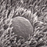
New microscopy method maps lipids without labels
A new microscopy method reads lipids' natural spectral fingerprints using mid-infrared light and ultrasound – no fluorescent labels needed, less stress for living cells.


A new microscopy method reads lipids' natural spectral fingerprints using mid-infrared light and ultrasound – no fluorescent labels needed, less stress for living cells.
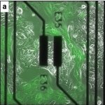
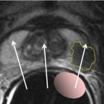
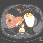
A new analytical method could improve how cancer treatments are designed – by allowing scientists to track, for the first time, exactly where inside a living cell a drug accumulates.
Bacterial infections can be difficult to distinguish from viral infections and inflammation, but a simple breath test may change that. This could reduce unnecessary antibiotic prescriptions.
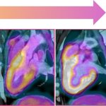
A widely used chemotherapy drugs used in cancer treatments can cause heart damage, new research shows. This could be used to adapt treatment regimens - especially in patients with high blood pressure.

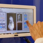
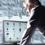
With antimicrobial resistance causing over 5 million deaths annually, rapid outbreak detection is critical. A German lab demonstrates how FTIR spectroscopy can transform hospital infection control.
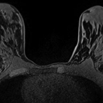
Dense calcifications are common in breast tissue, but not every finding in a mammogram is a precursor of cancer. New insights could lead to fewer benign biopsies and guide therapeutic development.
Through quantitative real-time monitoring of cell spatiotemporal dynamics, or cell changes over time, electrical impedance spectroscopy (EIS) could be used to predict cancer growth.
By identifying the unique biological signatures that guide AI tissue classification, new research lays the foundation for smart surgical tools with the added feature of biomolecular-level precision.

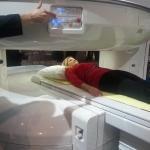
Breast MRI has emerged as a powerful diagnostic tool, particularly for women with dense breast tissue where traditional mammography faces limitations. In her presentation at ECR 2025, radiographer Hanna Kalliomäki highlighted several technological advances transforming breast cancer detection and diagnosis. From time-saving abbreviated protocols and AI-assisted analysis to contrast-free…
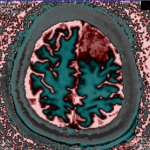
Blood-based biomarker (BBB) tests may represent the best weapon to combat the soaring rates of Alzheimer’s disease (AD) throughout the world. Existing clinically validated tests are currently deployed to facilitate diagnosis, to monitor disease and effectiveness of treatments, to quantify progression, and to determine if a patient is appropriate for treatment or participation in a clinical…
Through the joint efforts of United Imaging and NeuralMed, Prizren Regional Hospital has become the first facility in Kosovo to benefit from United Imaging's cutting-edge diagnostic technology.
Some year in this decade, AI tools will become ubiquitous within clinical laboratories. AI has the potential to increase the accuracy of laboratory testing and improve the quality and efficiency of operations and service of testing labs.
A new approach to the identification of harmful bacteria: A new study explores how spectroscopic techniques can be used for quick analysis directly from the skin.
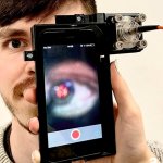
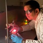
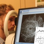
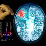
Researchers from the University of Birmingham have designed and developed a novel diagnostic device to detect traumatic brain injury (TBI) by shining a safe laser into the eye.
Cedars-Sinai Medical Center, a non-profit hospital and medical research institution in Los Angeles, is setting new standards for quality and innovation in patient care by successfully introducing typing of Candida auris species – a procedure that could prove crucial in protecting patients from infection outbreaks caused by these microbes in healthcare settings.

Under the impulse of the European Commission, the in vitro diagnostic industry is developing emerging technologies to implement sustainable practices in medical laboratories. As sustainability has been a growing priority of the European Union (EU) in the last decade, ‘the medical technology sector, particularly the IVD sector, must comply with European legislation in this field like all other…
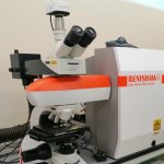
Researchers have developed a Raman microscope that can acquire information hundreds of times faster than the conventional method. This could help expand its usefulness in biomedical applications.

Monitoring the proper blood supply to the brain could be used to prevent or even treat neurological diseases. A new technique called πNIRS aims to do just that.
New method for the detection of alcohols combines zero- to ultralow-field nuclear magnetic resonance with the SABRE-Relay hyperpolarization technique.

Researchers are seeking alternatives to gadolinium-based contrast agents (GBCAs) to help raise levels of patient safety. An open hybrid session at ECR in Vienna heard how research across several centres has been examining the options of new approaches to reduce reliance on GBCAs.
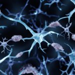
Copper exposure in the environment and the protein alpha-synuclein in the human brain could play an important role in the pathogenesis of Parkinson's disease, researchers found.
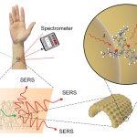
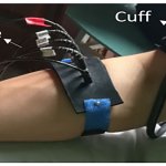
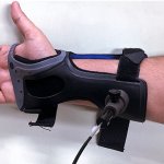
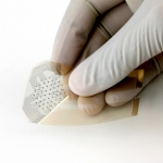
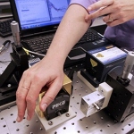
Researchers created a special ultrathin sensor, spun from gold, that can be attached directly to the skin without irritation or discomfort. The sensor can measure different biomarkers or substances to perform on-body chemical analysis.
Hyperpolarized nuclear magnetic resonance enables major medical advances in molecular diagnostics, for example for cardiovascular diseases or cancer therapy.

The advantages and limitations of the value of traceability chains in therapeutic drug monitoring will be explored during a key session at the EuroMedLab event.
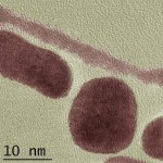
Researchers have succeeded in using metal oxide nanoparticles as "radiosensitizers" – and in producing them on an industrial scale.
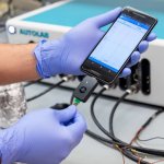
The tool has made it possible to detect SARS-CoV-2 in exudate from symptomatic patients with a sensitivity of 100% and a specificity of 87.5%
Researchers have developed a noninvasive and reagent-free technique for the efficient detection of COVID-19.

Researchers from Auckland have uncovered a link between the sharpness of surgical tools such as scalpels and the risk of post-surgery infection.
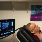
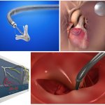
The EU-funded consortium Laser and Ultrasound Co-analyzer for Thyroid Nodules (LUCA) has developed a non-invasive, low-cost device that brings a new solution for thyroid cancer screening.
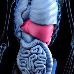
Handheld laser devices that help surgeons quickly spot liver damage could transform transplant procedures, research suggests. The non-invasive technique could provide medical staff with instant data on the health of donor livers and help them to identify which organs are suitable for transplant. If widely adopted, the light-based tool could allow more livers to be transplanted safely and…
A low-cost, rapid diagnostic test for Covid-19 developed by Penn Medicine provides Covid-19 results within four minutes with 90 percent accuracy. A paper published this week in Matter details the fast and inexpensive diagnostic test, called RAPID 1.0 (Real-time Accurate Portable Impedimetric Detection prototype 1.0). Compared to existing methods for Covid-19 detection, RAPID is inexpensive and…
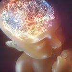
A new photonics device currently in development aims to reduce unnecessary disabilities by improving the instant, real-time monitoring of newborn babies with harmless light particles.
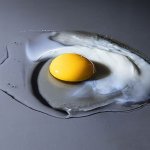
A team of scientists has been using the X-ray source PETRA III at the German Electron Synchrotron (DESY) to analyse the structural changes that take place in an egg when you cook it. The work reveals how the proteins in the white of a chicken egg unfold and cross-link with each other to form a solid structure when heated. Their innovative method can be of interest to the food industry as well as…
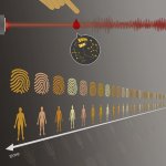
A new study carried out by a team of laser physicists, molecular biologists and physicians based at LMU Munich and the Max Planck Institute for Quantum Optics has confirmed the temporal stability of the molecular composition of blood in a population of healthy individuals. The data provide a basis for a new method of monitoring the constituents of blood and detecting alterations that reveal…
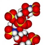
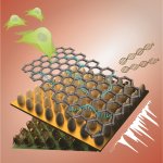
Using a nanopore, researchers have demonstrated the potential to reduce the time required for sequencing a glycosaminoglycan — a class of long chain-linked sugar molecules as important to our biology as DNA — from years to minutes. Research to be published this week in the Proceedings of the National Academies of Sciences shows that machine-learning and image recognition software could be…
Improving the detection of cancerous cells during surgery – this is the goal of the European research project CARMEN. The research institutes Laser Zentrum Hannover e.V. (LZH) from Germany and Multitel asbl from Belgium work together with companies from both countries, JenLab GmbH, Deltatec, and LaserSpec, to develop a novel, compact and multimodal imaging system. This could even allow the…

Increased consumption of flavanols – a group of molecules which occur naturally in fruit and vegetables – can increase your mental agility, according to new research. A team at the University of Birmingham has found that people given a cocoa drink containing high levels of flavanols were able to complete certain cognitive tasks more efficiently than when drinking a non-flavanol enriched-drink.
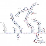
For the first time, an international research alliance has observed the RNA folding structures of the SARS-CoV2 genome with which the virus controls the infection process. This could not only lay the foundation for the targeted development of novel drugs for treating Covid-19, but also for occurrences of infection with new corona viruses that may develop in the future.
University of Maryland scientists have developed a method to determine the structures of large RNA molecules at high resolution.

New techniques of infrared-based technology are showing strong potential for cost-effective tissue analysis. Peter Gardner, Professor of Analytical and Biomedical Spectroscopy at the University of Manchester, outlined how hyperspectral imaging coupled with sophisticated computer algorithms can identify and grade cancerous tissue, as well as offer an indication of prognosis. The technique, he…
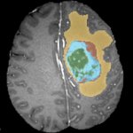
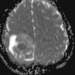
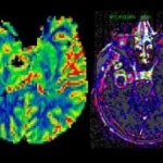
Neuroradiologist Dr Sofie Van Cauter described the challenges to brain tumour image segmentation during the European Society of Medical Imaging Informatics (EuSoMII) annual meeting in Valencia. She also outlined how, when clinically validated, AI could help tackle such problems. The WHO classification of brain tumours has come a long way since first introduced in 1979. The 2016 classification was…

In diagnostics, it sometimes makes sense to follow your nose. During the Labmed Forum at Medica, Dr Beniam Ghebremedhin and Dr Simona Cristescu discussed the diagnostic potential of breathomics – the analysis of a patient’s exhaled air for disease indicators.
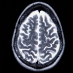
A simple blood test coupled with artificial intelligence (AI) analysis could help spot the signs of a brain tumour sooner in patients. Brain tumour diagnosis is difficult: patients often see their family doctor (GP) several times before referral for a scan. However, research presented at the 2019 National Cancer Research Institute (NCRI) Cancer Conference in Glasgow last November suggests the…

Ground-breaking research by the University of Birmingham has discovered a new technique to assess the aggressiveness of childhood brain tumours. Funded by Children with Cancer UK, Action Medical Research and The Brain Tumour Charity, the study is the first of its kind and will allow clinicians to give more personalised treatments for childhood brain cancers, which currently account for one third…
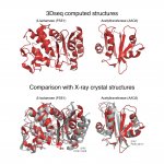
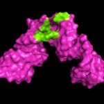
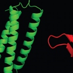
“Proteins are the workers in the cell, and it's important to know their shape,” says Chris Sander, PhD, director of Dana-Farber’s cBio Center in the Department of Data Sciences. Sander and his colleagues have now demonstrated a powerful “experimental evolution” method to discover details of protein shape and function, and the method may find uses across a very broad spectrum of…
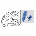
Parkinson’s and multisystem atrophy (MSA) – both of them neurodegenerative diseases – are associated with the accumulation of alpha-synuclein proteins in the brain. Researchers at the German Center for Neurodegenerative Diseases (DZNE) and the Max Planck Institute for Biophysical Chemistry (MPI-BPC) have investigated the molecular makeup of these protein deposits finding structural…
Researchers have created better biosensor technology that may help lead to safe stem cell therapies for treating Alzheimer’s and Parkinson’s diseases and other neurological disorders.
Early non-invasive detection of kidney rejection after transplantation was the central aim of a collaboration between Prof. Dr. Bernhard Banas, Chairman of Nephrology at the University Hospital Regensburg (UKR) and the medical diagnostics company, numares. The results of their joint clinical trial “UMBRELLA” were just published in EBioMedicine and presented at the American Society of…
Chemical analysis of blood samples, combined with an artificial intelligence program, could speed up the diagnosis of brain tumours, according to research presented at the 2019 NCRI Cancer Conference. Brain tumours tend to have ambiguous symptoms, such as headache or memory problems, and a brain scan is currently the only reliable way of diagnosing them. Researchers say their test, which works by…
More and more people aged 50 and over are suffering from age-related vision disorders. According to the World Health Organization, in four out of five cases they could be avoided if they were diagnosed at an early stage. A European team of scientists, including the Leibniz Institute of Photonic Technology (Leibniz IPHT) in Jena, has now researched a new method that will enable doctors to better…
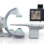
Interventional cardiologists have been aware of the value of intracoronary (IC) imaging in clinical practice for more than twenty years. However, recent developments and improvements in modalities and software have enabled huge strides in its range and scope for both diagnostic assessment and in percutaneous coronary interventions. Imaging options now include intravascular ultrasound (IVUS),…
A remarkable number of studies and innovations were presented at the 30th anniversary of Transcatheter Cardiovascular Therapeutics (TCT) meeting in San Diego, California. TCT 2019 will take place in San Francisco, CA between 25-29-Sep-2019. On the clinical side, the long-expected results from COAPT trial studying MitraClip device in patients with secondary mitral regurgitation and heart failure…
Researchers from Northwestern University are using Argonne supercomputers to advance the development of an optical microscopy technique that can predict and quantify cancer risks at extremely early stages. The basic principle driving Allen Taflove’s computational electrodynamics research — which bears the potential to transform how we diagnose, and possibly treat, various forms of cancer —…
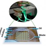
Analytically sensitive and specific detection of pharmaceuticals or metabolites in bodily fluids, as well as fast and reliable detection of human pathogens, are major challenges for instrument-based analytics in medical diagnostics. Over the past few years the combination of surface-enhanced Raman spectroscopy (SERS) and microfluidic devices (Lab-on-a-Chip) has emerged as a perfectly suited…
A new way of detecting rheumatoid arthritis using infrared light could offer an objective way of diagnosing the disease and monitoring treatment effectiveness, a University of Birmingham study shows. The rapid, non-invasive technique could help clinicians diagnose the disease earlier, and assess how effectively the selected treatment is controlling the progression of the disease. Rheumatoid…
Cheap and compact instrument based on two near-infrared light techniques diagnoses poor blood flow quickly and noninvasively.
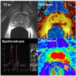
Pathologists, radiologists, urologists and radiotherapy specialists sit at the core of the treatment pathway for the patient, working together as a cohesive unit. In an innovative 2019 ECR session in Vienna, the prostate unit from Ghent University Hospital in Belgium outlined how the team works to deliver the best clinical outcome for patients.
Scientists at the University of Birmingham are working with a Canadian tech company to investigate whether gold nanorods can be used to target cancer cells in the human body. They have joined experts at Sona Nanotech Inc. to develop the next generation of nanorods for tissue imaging. The team will work with its Canadian partners - beginning by creating luminescent nanorods by transforming gold…

As Shimadzu celebrates its 50th anniversary in Europe, we spoke with Stéphane Moreau, Manager of LC-MS & Life Sciences at the Marketing Europe/Analytical Business Unit of Shimadzu Europa GmbH, about today’s and many more decades of exciting clinical developments.
Researchers recently evaluated the accuracy of a technology to monitor blood glucose levels without needles or a finger prick. Early results show that the noninvasive technology measures blood glucose levels as effectively as a finger prick test — without drawing blood.
Scientists at the Virginia Tech Carilion Research Institute (VTCRI) have found evidence that may disrupt conventional understanding about how electrical activity travels in the heart — a discovery that potentially can lead to new insight into medical problems such as heart arrhythmia and sudden cardiac death.
MRI has developed rapidly over the past decade in Poland, where clinicians are combining MRI with PET and CT to highlight tumour growth or regression and perfusion. ‘The fact that MRI offers new software and programmes means we can diagnose pathologies more precisely and make a diagnosis faster than a few years ago,’ explained Poland’s national advisor on radiology and diagnostic imaging…
Gadolinium-containing/gadolinium-based contrast agents (GCCAs/GBCAs) and their usage was a major topic at ECR 2018. Fuelled by the current debate a number of presentations focused on possible impact, risks and necessities. Some were highly specific, others took a broader view. The only consensus, however, seems to be the need for more research and the focus on safety. Three ECR speakers, Joseph…
Engineering researchers at the University of Arkansas have moved closer to developing an alternative method of detecting and possibly treating breast cancer. Terahertz images of breast tumors taken from mice accurately detected cancerous tissue with a high level of specificity.
Recognising malignant tissue remains a tricky task. While today, most patients undergo a biopsy, an invasive procedure where tissue is sampled, stained and assessed, researchers are exploring the potential of optical biopsy, the visual assessment of suspect tissue. The interest in optical biopsy ‘is indeed enormous,’ confirms Dr Thomas Bocklitz, physicist at Friedrich-Schiller University in…
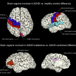
The emerging field of psychoradiology is taking a major step ahead. A new study highlights MRI’s role in identifying people with attention deficit and hyperactivity disorder (ADHD) and classifies subtypes of the condition, a leading Chinese researcher explained at the ESMRMB annual meeting.

Breast diagnostics are undergoing considerable change, with new technology facilitating alternative procedures. Genetics and nuclear medicine also enhance diagnostic possibilities. During our EH interview, Professor Rüdiger Schulz-Wendtland described current changes in breast diagnostics. ‘To date, complementary breast diagnostics has comprised clinical, sonographic and mammographic…
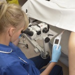
A pioneering diagnostic device identifies more abnormal cells on the cervix than standard colposcopy, according to data published in two European clinical papers.
Canadian researchers have invented an intraoperative probe that reliably detects multiple types of tumour cells.
Current advances such as phase-contrast CT are taking medical imaging further but their use in clinical practice may have to wait up to a decade, prominent physicist predicts.

The recipe for an exciting congressional session is simple: Take a controversial hypothesis, a proponent who defends this hypothesis and an opponent who argues against it.

From the extremely new, but not generally available, to the somewhat new… very available and highly useful… Walter Kucharczyk outlines potentials and practicalities in advanced brain tumour Imaging.
From the extremely new, but not very available, to the somewhat new, very available and highly useful, Walter Kucharczyk will cover the potentials and practicalities in advanced brain tumor imaging.
Pathology does not appear to have much in common with satellites, but the concept that satellites combine spatial resolution and image quality will be the future of disease diagnosis, according to researchers.

From barcode scanning, RFID and medical machine vision to chart recorders, thermal printers and spectroscopy products, JADAK designs and manufactures embedded solutions that help customers solve unique inspection, tracking, scanning and documenting challenges.
UK researchers are working on a more precise imaging technique for dilated cardiomyopathy that may lead to more effective treatments. A study from the University of Oxford Centre for Clinical Magnetic Resonance Research (OCMR), part of the Division of Cardiovascular Medicine at the university, has demonstrated how the next generation of MRI scanners can work to measure heart conditions in dilated…

Photo Research (PR) has merged with its Novanta sister company JADAK, a manufacturer of machine vision, radio-frequency identification (RFID) and bar code products for the health care and life science industries. Photo Research is a leader in world-class light and color measurement solutions serving the flat panel display, automotive, aerospace and related industries.
The unusual electronic properties of some superconducting materials permit lossless and dense electrical currents at very low temperatures, even in high magnetic fields. Conductors made of these materials are thus ideal for winding coils to generate very high magnetic fields, which are essential for a number of applications like magnetic medical imaging, MR spectroscopy for the analysis of…
New, screen-printed, flexible MRI coils may be able to reduce the amount of time it takes to get an MRI scan. Researchers funded by the National Institute of Biomedical Imaging and Bioengineering (NIBIB), part of the National Institutes of Health (NIH), have developed light and flexible MRI coils that produce high quality MRI images and in the future could lead to shorter MRI scan time periods.
As a leading global provider of both diagnostic imaging and analytical instrumentation technologies, Shimadzu offers broad expertise in medical imaging and mass spectrometry detection platforms helping to deliver a measurable impact on healthcare and diagnosis. The company is the perfect partner for transformational technologies to accelerate diagnosis.
‘In imaging there is a trend towards quantification,’ said Professor Siegfried Trattnig, Medical Director of the High-Field MR Centre (HFMRC) at the Medical University Vienna, Austria. Whilst before, radiologists’ findings were subjective, qualitative results, based on signal intensity and grey scale, he pointed out. ‘Today imaging can draw on quantifiable and comparable parameters with…

The detection of light by pigments in the retina, called rhodopsin or visual purple, leads to our sense of vision. New experiments by scientists from the Max Planck Institute for the Structure and Dynamics of Matter and the University of Toronto have revealed that the primary photochemical event of this process operates at the fundamental molecular speed limit.
Engineers at the University of California, Berkeley, are developing a new type of bandage. Thanks to advances in flexible electronics, the researchers have created a new "smart bandage" that uses electrical currents to detect early tissue damage from pressure ulcers, or bedsores, before they can be seen by human eyes - and while recovery is still possible.
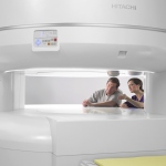
As Hitachi deploys its new generation of Oasis 1.2-T MRI scanners throughout Europe, our Madrid correspondent asked Dr Manuela Jorquera Moya about her experiences with the new scanner over the past few months.
Researchers at the Virginia Bioinformatics Institute at Virginia Tech have uncovered key cellular functions that help regulate inflammation -- a discovery that could have important implications for the treatment of allergies, heart disease, and certain forms of cancer.

The field is neither tedious nor monotonous; it’s fascinating every day, Professor Katharina Rentsch emphasises, when explaining the need to attract students to this often overlooked but intreguing and varied discipline.
Brain lesions in children can be especially challenging to diagnose, according to a report in the journal Frontiers in Neurology by a multidisciplinary team of Loyola University Chicago Stritch School of Medicine physicians. Lesions include tumors, abnormal blood vessel formations, and abscesses and inflammation due to infections.
Hitachi has double-down its bet on the advantages of open-platform MRI by introducing a new generation of the Oasis 1.2T scanner this year at RSNA. The jump to new processing power and the new Origin 4.0 MR Operating Software effectively enhances capabilities with a range of new applications for neuro, orthopedic and vascular imaging as well as enhancing routine exams for women's health and…

Scientists who use synchrotron light sources are welcoming an era of "on-demand" X-rays, in which they have access to the light beams they want thanks to a technique developed at the U.S. Department of Energy (DOE)'s Lawrence Berkeley National Laboratory (Berkeley Lab).

In breast cancer care each patient receives personalised, highly effective diagnosis and treatment procedures. In breast diagnostics this mainly revolves around new MRI scanning procedures that allow the quantification of biological and physiological processes on a cellular and molecular level.
Integration of imaging biomarkers in Europe is a challenge, and a number of issues must be overcome so that the full potential of quantitative imaging can be realised, renowned researchers will show in a session today at the ECR.

The son of a craftsman making Buddhist altars, he was driven to create instruments for physics and chemistry. Attending the Physics and Chemistry Research Institute he gained experience with a variety of technologies and fields of expertise. He was convinced that Japan, as a should work towards becoming a leader in science. At the dawn of the industrial revolution and scientific age in 1875 he…
A magnetic resonance spectroscopy (MRS) technique that monitors biochemical changes in tissue could improve the management of women at risk of breast cancer.
Glucose testing is both a headache and an opportunity for clinical laboratories here in the United States and across the globe. It is a headache because many point-of-care and patient self-test glucose devices in wide use today lack the reliability of glucose testing performed in medical laboratories that use sophisticated diagnostic instruments.

Refined acquisition techniques and coils facilitate the assessment of cranial nerves with MRI. Professor Dr Elke Gizewski, Director of the University Clinic for Neuroradiology at the Medical University Innsbruck, Austria, is an expert in diagnostic and interventional neuroradiology and explains pathologies and scanning techniques for intracranial nerves.
The human gut literally teems with microorganisms from at least 1,000 different species that are increasingly considered to be a valuable resource for the prediction, aetiology and prognosis of disease. Due to continual contact with the environment, primarily via food, the gut is susceptible to infection when a virus, parasite or bacterium enters and disrupts normal gut microbiota (or flora).
Although telemedicine could improve the quality of life of patients with chronic liver diseases, viable home care systems are still lacking. However, within the EU-project ‘d-LIVER’ (www.d-liver.eu) scientists at the Fraunhofer Institute for Biomedical Engineering IBMT, in St. Ingbert, Germany, are working with European partners to develop an IT- and cell-based system that will help chronic…
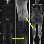
With MRI and CT scanners widely available in clinical routine, radiologists cull increasingly precise and relevant functional tumour information for diagnostics and monitoring purposes. Both modalities offer technological and methodological approaches, initiated by the discipline itself, that have become indispensable for certain frequent tumours.
Medical technologies have maintained a leading position in European patent registrations for 15 years.

Quality management (QM) for medical laboratories shaped its footprint during recent years – it’s a hot topic in papers, conferences and trade fairs. A lot of information makes a lot of work, says Dr Markus Neumann
Medicine is not immune to prejudices. In the past, the ‘fatty liver’ diagnosis was often accompanied by the hasty conclusion that the problem was surely caused by alcohol abuse.

Hedvig Hricak, Chair of the Radiology Department at the Memorial Sloan-Kettering Cancer Center, New York, USA, describes emerging applications and potential trends in gynaecological cancer treatment described at the 15th International Symposium Crossing Barriers

Photoacoustics offers an additional contrast mechanism that makes the technology far more sensitive for the presence of blood.
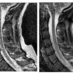
The fact that between 60% and 80% of people are expected experience some form of back pain at some point in their lives puts the importance of advances in imaging of the spine into context. A number of developments in imaging of the spine and peripheral nerves were outlined at a musculoskeletal scientific session at ECR 2012 in Vienna on Saturday morning.
Radiology constantly evolves. There are technical advances in terms of the capabilities of various modalities, greater clarity from contrast agents that are also safer for patients, and innovation in techniques that gains even greater performance from existing equipment, or enables further development.
As a referral neuroradiologist for paediatric tumour studies, Professor Monika Warmuth-Metz, Consultant at the Neuroradiology Department at University Hospital Würzburg, daily evaluates MRI images of different origin and colour. Her resume states: ‘All too often the standard protocols set out in the guidelines are not adhered to, which makes evaluation and follow-up significantly more…
Vienna - For the 23rd time, the European Congress of Radiology (ECR) is opening its doors to welcome 19,000 participants from over 90 countries. The scientific exchange of knowledge and the presentation of the latest developments in the field of radiology will again be presented right in the heart of Europea. In an inaugural press conference on March 3rd, the hot topics of the congress were…
Thinking of the future of imaging, inevitably PET-MRI springs to mind. The fascination of this novel hybrid technology is great, seeing how it combines the best from three imaging areas: anatomy, function and metabolism. The further development of functional procedures in oncology is raising particularly high expectations. However, how extensive the use of this potentiated image information will…
Muscular diseases belong to a heterogeneous group with various causes like neurogenic, metabolic, dystrophic, or inflammatory mechanisms as well as channelopathies leading to disorders of the muscle cell membrane potential. In most progressive disease cases the result is a focal or general muscle weakness that, unfortunately, is a very unspecific symptom. Standard neuromuscular literature…
Early problems of ultra-high field Magnetic Resonance Imaging (MRI) have been overcome by successful development of adequate hardware. In consequence big efforts have been achieved in structural imaging, as well in functional imaging. Basic scientists and physicians who work in ultra-high-field MRI in Europe and the USA, met at the Berlin Ultra-high-field Facility (BUFF), in the Max Dehlbrück…
Intra-operative MR imaging is still uncommon in Europe – particularly in neurosurgery. One of the first neurosurgery departments to work with a 3-T MR is in the Central Military Hospital in Prague. The experience and initial clinical findings from treating more than 300 patients, over a two-year period, was reported at this year’s ECR.

Molecular imaging, a new science that emerged from molecular biology, is unlike traditional imaging. Whilst the latter can, for example, show the differences in proton density or water content on MRI, molecular imaging uses biomarkers (probes) that interact selectively with molecules within an area and then generate the image according to fine molecular alterations occurring inside (e.g. within a…
The technological integration of positron emission tomography (PET) and magnetic resonance imaging (MRI) has been the dream of molecular imaging experts and engineers for some time. Now, the German Science Council has agreed to provide 6.56 million funding to install a whole-body MRI-PET prototype in the centre of excellence for imaging procedures at the radiology clinic in Eberhard-Karls…
When we organised the first Diagnostic Week in Karlsruhe, in 1969, no one could have known that this event would one day turn into the annual highlight in the world of medicine, reflected Dr Wolfgang Albath, laboratory medicine pioneer and one of the founding fathers of MEDICA the world`s largest medical trade show. Initially planned as a moving exhibition, the show has been based in…

For the past 30 years, in the former USSR and Russia, laser medicine has been actively developed and regular annual conferences have opened new avenues for its use by the country's doctors.

Emphasising the crucial partnership of radiologists and urologists in the treatment of prostate cancer, Professor Arnulf Stenzl, Medical Director of Urology at the Tübingen University Hospital, explained that, throughout the phases of prostate cancer diagnosis and therapy - from primary diagnosis onward - imaging is indispensable.
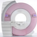
On this years ECR Siemens Healthcare presents its dedicated MRI breast scanner. With it's pink design it immediately catches the eye on the company's booth. In an interview with EH, Boris Tolkien, Vice President Marketing Magnetic Resonance, Siemens Healthcare pointed out the highlights of the 1.5 Tesla system beyond its colour.
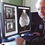
Dr Giorgio Rizzatto, who heads the Department of Diagnostic Imaging at ASS 2 Isontina (including the Gorizia and Monfalcone hospitals) in NE Italy, works with nine radiologists, who examine and report on 80,000 clinical and screening breast examinations annually.

Big discussions are expected at the upcoming European Association of Urology (EAU) Congress in Stockholm* when urologist Dr Jochen Walz (right), of the Urological Department at the Institute Paoli-Calmettes, Marseilles, France, presents the forum: Imaging in Europe: Who, where, what, how many! and M F Coelho, of the European Society of Urological Imaging (ESUI) describes the Clinical utility of…
The machine 'qViro' which is the size of a coffee grinder will make virus counting much cheaper than traditional virus counting technology. The revolutionary new invention offers the potential to quickly muster and count the number of viruses in a sample in minutes - it is a portable, desktop instrument, powered from the USB drive of a computer - current competitors are the size of washing…
The ratio between the concentrations of metabolites may give the answer to the question. Researchers at the Helmholtz Zentrum in Munich demonstrated the proof of principle. They identified diabetic or healthy mice by biomarkers they analyzed only by bioinformatics. Metabolomics might permit a promising tool for pre-clinical investigation of effects and side effects of new drugs, they say.
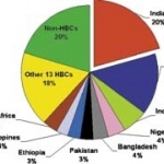
Every three minutes, two people living in India die of tuberculosis. This equates to approximately 370,000 deaths each year, and a staggering economic toll: an estimated US$300 million in direct costs and US$3 billion in indirect costs.

Prostate cancer is the most frequent cancer in men with about 49,000 newly diagnosed cancers in Germany and 6,000 in the Netherlands, annually.
Only few imaging modalities lend themselves to imaging of the lungs. Conventional chest radiography is the most commonly used tool in the investigation of pulmonary pathology but yields the perhaps most difficult, plain radiographs to interpret.
A team of Stanford University School of Medicine researchers has developed a new type of imaging system that can illuminate tumors in living subjects-getting pictures with a precision of nearly one-trillionth of a meter.
Can they replace PET? By Marco Essig MD, Professor of Radiology at the German Cancer Research Centre, outlines relevant presentations at the ECR.

For individualised radiotherapy, high-precision delineation and characterisation of the tumour is critical. If highest radiation doses are delivered in a targeted fashion, the chance of tumour cell kill increases and tumour control probability is enhanced.
The Uliazpi Foundation in Spain, which studies and cares for severely mentally retarded patients, carried out an interesting study to identify bone mineral density values in a group of its patients, compare these with the general population and investigate the possible influence on these values on certain clinical variables and therapeutic regimens.
As reported in the January issue of Hepatology, MRI imagery is emerging as a non-invasive way to determine the existence and extent of hepatic fibrosis. It could eventually help the development of pharmacologic strategies to combat the condition.

USA - MRI is playing an increasingly important role in breast cancer screening in the US, especially in the screening of high risk patients. It has proven to be more sensitive than mammography examinations, but is less accurate in the differentiation between benign and malignant lesions.

Presented during a symposium at Langenbeck-Virchow-Haus, in Berlin, the European CLINICIP research project aims to develop a method to improve glycaemic control during intensive care and provide a low-risk monitoring and control system that can control the metabolism of the critically ill
IVAM is an international association of companies and institutes that specialise in microtechnology, nanotechnology and advanced materials.
Hutchinson's InSpectra StO2 Tissue Oxygenation Monitor - a portable device for treatment of trauma patients.
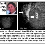
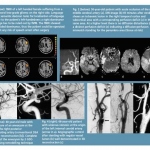
By Stefan G Ruehm MD PhD, Associate Professor of Radiology, at the David Geffen School of Medicine, UCLA, California
As the German Society of Neuroradiology 40th annual meeting approached (Venue: Dresden. 31 August - 3 September), Professors Martin Schumacher (Freiburg), President of the German Society of Neuroradiology (GSN) and Rüdiger von Kummer (Dresden), the meeting's President, examine the history and potential in this medical field