Photo: Ewald Unger/ Medical University of Vienna
News • Macular degeneration as a biomarker
Eye scan shows diseases at an early stage
More and more people aged 50 and over are suffering from age-related vision disorders. According to the World Health Organization, in four out of five cases they could be avoided if they were diagnosed at an early stage.
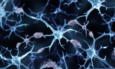
A European team of scientists, including the Leibniz Institute of Photonic Technology (Leibniz IPHT) in Jena, has now researched a new method that will enable doctors to better detect such eye diseases in the future. The optical method uses laser light to provide detailed information on the condition of the retinal tissue in a matter of seconds. With this eye scan, physicians will be able to detect aggressive forms of age-related macular degeneration sooner and even detect neurodegenerative diseases such as Alzheimer's disease.
Clara Stiebing and the research team from Leibniz IPHT, Friedrich Schiller University Jena, Medical University Vienna and partners from the Netherlands published their study in the journal "Neurophotonics".
Recommended article
Article • Surface-enhanced Raman spectroscopy
The lab-on-a-chip SERS platform
Analytically sensitive and specific detection of pharmaceuticals or metabolites in bodily fluids, as well as fast and reliable detection of human pathogens, are major challenges for instrument-based analytics in medical diagnostics. Over the past few years the combination of surface-enhanced Raman spectroscopy (SERS) and microfluidic devices (Lab-on-a-Chip) has emerged as a perfectly suited…
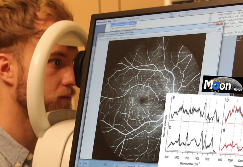
A laser beam hits the eye. This sounds more like a risk of injury at first, but in this case it opens up a chance of healing. "We use laser light to obtain comprehensive molecular information about the retina and thus early indications of diseases," explains Stiebing. In order to find out how much laser power the eye can tolerate and which optical pathway the laser takes, the researcher examined retinal samples and designed a structure simulating the conditions in the human eye.
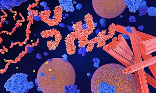
How intense the laser can be without harming the eye, the scientists have calculated exactly on the basis of applicable safety regulations. The result: a laser beam that is twenty times weaker than the lasers that researchers otherwise use for their spectroscopic measurements. Using label-free, molecularly sensitive Raman spectroscopy, they are able to obtain a molecular fingerprint of the retina. This reveals how high the content of lipids, proteins, carotenoids and nucleic acids is. In this way, changes in the retina become visible, enabling doctors to detect diseases at an early stage.
The fact that we can now supplement the OCT previously used in ophthalmology with Raman spectroscopy can significantly improve the accuracy of the diagnoses
Jürgen Popp
A particular challenge for the researchers was that the conditions in the human eye were not optimal for optical measurements. "The fact that we are still able to achieve meaningful, reliable results with the attenuated laser beam clearly shows that our technology will enable us to obtain comprehensive molecular information about the structure of the retina in the future," said Clara Stiebing. The partners of the Medical University of Vienna are currently building a device that combines Raman spectroscopy with optical coherence tomography (OCT). With the help of OCT, the morphology of the retina can be very quickly made visible and suspicious areas can be identified. Raman spectroscopy can then be used to characterise these areas at the molecular level. "This enables us to obtain high-resolution images from all layers of the retina including information on their molecular composition," explains Prof. Jürgen Popp, scientific director of Leibniz IPHT. "The fact that we can now supplement the OCT previously used in ophthalmology with Raman spectroscopy can significantly improve the accuracy of the diagnoses.“
The European research team is currently working on the medical approval of the device. Once approved, the device can be tested on patients. The patients would then sit in front of the device, have their eye scanned without contact and a few minutes later, they would receive a reliable diagnosis.
Source: Leibniz Institute of Photonic Technology (Leibniz IPHT)
06.09.2019