ECR 2011 prelude
Vienna - For the 23rd time, the European Congress of Radiology (ECR) is opening its doors to welcome 19,000 participants from over 90 countries. The scientific exchange of knowledge and the presentation of the latest developments in the field of radiology will again be presented right in the heart of Europea. In an inaugural press conference on March 3rd, the hot topics of the congress were revealed.
ESR President Prof. Dr. Maximilian Reiser, Dean of the Medical Faculty at the Ludwig-Maximilians University, Munich, Germany, opened the press conference reporting on the contribution of radiology to personalised medicine and cost containment in healthcare. “Over recent years and decades we have witnessed the steady increase in the importance of radiology to the healthcare system. Thanks to crucial developments since 1975, imaging has become more indispensable than ever to accurate diagnosis, which is a prerequisite for appropriate treatment,” he said. It is estimated that imaging now contributes to 70 to 80% of all diagnoses made in clinical medicine.
The more accurate the imaging modality, the earlier diseases can be detected, which in turn improves treatment outcome while reducing its costs. In breast cancer, detection of the tumour before metastatic spread means breast-conserving surgery may still be carried out, while no adjuvant chemotherapy is required. Identification of a poor response to a particular therapy means doctors can abandon a treatment option that may be costly and have associated side effects.
In order to fully exploit the potential of radiological imaging and intervention to contain healthcare costs, radiological practice has to be based on scientific evidence, explained Reiser. “The appropriate method of imaging has to be applied and high standards of training have to be provided. Moreover, standardised reporting must guarantee that the referring physicians obtain the information required for the management of their patients,” he said.
Meetings must reflect new trends in medicine
ECR 2011 Congress President Yves Menu, Chairman of the Dept. of Radiology, Saint Antoine Hospital, Paris, France, took on business by focusing on the future of meetings, emphasizing: “Making a congress today cannot be the same as it was decades ago,”
Subspecialisation has increased, making communication more difficult between different subspecialists within a discipline like radiology, he explained. The range of knowledge has broadened considerably, with a huge number of imaging tools that did not exist thirty years ago. And while e-communication has improved immeasurably, financial limitations in times of crisis and ecological concerns restrain the desire or ability to travel.
All these factors call for a change in the way international conferences are being held.
“One idea would be just to cancel all meetings and organise them remotely, taking advantage of internet communication. However this would not take into account one of the main advantages of meetings; face-to-face communication,” Menu said. Experts are leaning towards transforming meetings from the inside, by taking into account new trends in modern medicine. Interactivity, personalisation and multidisciplinarity must be reflected in radiological congresses, he closed.
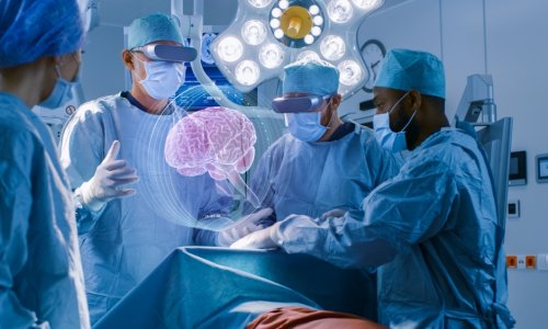
Clinical potential of molecular imaging: it's already reality!
Eventhough clinicians from every discipline acknowledge that molecular imaging will play a crucial role in personalised medicine and molecular therapy, especially in cancer management, nevertheless many are still put off by stereotypes, knows Fabian Kiessling, who since 2008 leads the Institute of Experimental Molecular Imaging at the Helmholtz Center of Applied Engineering of the RWTH-University in Aachen, Germany: “Many clinicians see molecular imaging as a discipline of ‘mouse doctors’ working in a highly artificial environment. One reason might be that clinical translation did not occur as rapidly as assumed. ”
Furthermore, molecular imaging tools are not recognised as such when they first enter the clinics, according to Kiessling. Prominent examples are MR spectroscopy and MR liver contrast agents such as SPIO or Gd-EOB-DTPA, the latter accumulating specifically in hepatocytes via scavenger receptors.
Molecular imaging applications are more prevalent in nuclear medicine. Many promising radiotracers that enable visualisation of different metabolic, physiological and molecular characteristics have been developed for PET and SPECT.
Unfortunately, the cost-intensive hurdles for clinical approval and the lack of sufficient radiochemistry facilities in the clinics often make pharmaceutical companies hesitant to invest heavily in the field, Professor Kiessling believes. “They prefer to focus on few broad ‘blockbuster pharmaceutics’ like Gd-DTPA,” he said.
In spite of difficulties, the field is advancing. New concepts for the synthesis and commercialisation of radiopharmaceuticals are currently being developed, and hybrid imaging modalities have seen molecular imaging become integrated with traditional radiological diagnostic methods.
7 Tesla at the MR Centre of Excellence Vienna
A 7 Tesla whole-body system with multinuclear capability is the flagship system of the MR Centre of Excellence (MRCE) of the Department of Radiology at the Medical University of Vienna. “The goal is to validate the potential of ultra-high-field MR in clinical applications,” said Professor Siegfried Trattnig, Head of the MRCE.
7T scanners are progressively becoming more widely available in Europe but there are only a few centres with 7T systems that are as close to an actual hospital as the MRCE. With the Vienna General Hospital (AKH), one of the largest hospitals in Europe, right next door, the MRCE is ideally positioned to carry out clinical research.
Ultra-short TE sequences open up the possibility to actually get signals from tissues like bone and tendons, which give no signal in normal MR imaging, meaning they just appear black in images. This means it is now possible to judge, for example, the internal structure of tendons, which could be an advantage in the early diagnosis of tendon damage. CEST is a method which enables selective visualisation and quantification of a constituent of cartilage or a spinal disc, namely a proteoglycan. This could be significant in the early diagnosis of cartilage damage, i.e. joint arthrosis, and spinal disc degeneration. This method could be used clinically in cartilage transplant patients for the first time in Vienna.
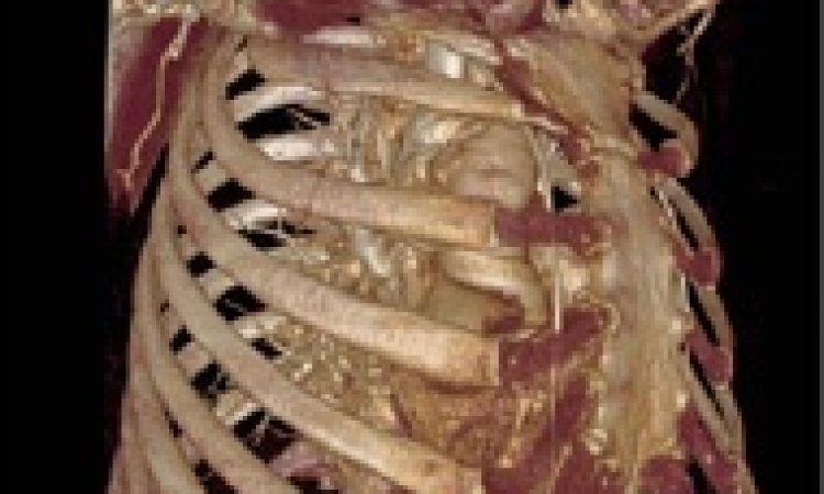
Slicing through works of art: CT leaks antique secrets
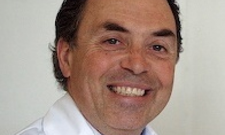
Computed tomography has been used to explore human bodies to help identify the origins of their pathologies since the late 1970s, so it is not surprising to find this modality occasionally used to probe reproductions of the human figure, explained Doctor Marc Ghysels from Brussels. He is an ex-interventional radiologist who plies his trade outside healthcare, using his skills to examine antiques and artworks from all over the world, and will present the Guest Lecture ‘Slicing through antiques and works of art’ on Saturday afternoon.
“Knowledge of human anatomy is irrelevant in this case, but most sculptures pass easily through any CT scanner, generating three-dimensional images which literally ‘undress’ the art work and reveal its internal structures and secrets,” said Dr. Ghysels.Radiological analysis of those internal structures may provide relevant information about the history of a piece, by revealing how it was made, what damage it has suffered over the years, how much restoration it has undergone, etc.
“Furthermore it can expose the many tricks used by forgers to deceive not only the discerning eye of the museum curator or collector, but also the more commonly used methods of scientific analysis,” Dr. Ghysels pointed out.
“Any radiologist with a bit of curiosity and access to a multidetector CT scanner can easily use his radiological skills to further our technical and cultural knowledge about art and antiquities, and about terracotta, wood and stone statues in particular. This is an atypical application for radiology which, although it is not new, has great potential for development as the number of authentic artworks now being found is dwindling every day and copies or fakes are invading the art market, whether in Chinese antiquities, African art or pre-Columbian terracotta,” Dr. Ghysels summarized.
03.03.2011