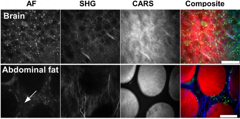
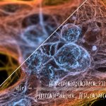
Image source: LZH (Graphic from J. of Medical Imaging, 2(1), 016003 (2015))
News • CARS & multiphoton microscopy
Multimodal imaging to detect cancerous cells faster and more accurately
Improving the detection of cancerous cells during surgery – this is the goal of the European research project CARMEN.
The research institutes Laser Zentrum Hannover e.V. (LZH) from Germany and Multitel asbl from Belgium work together with companies from both countries, JenLab GmbH, Deltatec, and LaserSpec, to develop a novel, compact and multimodal imaging system. This could even allow the examination of tissue samples directly during surgery.
Laser-based microscopes usually use only one imaging method, such as confocal microscopy, multi-photon microscopy or Anti-Stokes Raman Spectroscopy (CARS). Combining different imaging techniques in a single device makes it possible to obtain faster, more, and more reliable information about tissues and possible diseases. However, the various excitation lasers needed would make a complete system very complex, bulky, and expensive.
The partners in the CARMEN project want to develop an innovative laser system that generates several excitation wavelengths and different pulse durations. This would allow them to combine CARS with multi-photon and super-resolution STED (Stimulated Emission Depletion) microscopy in one compact device. Such a complete system would make it possible to examine tissue samples directly after the surgery or even during it. This would help to recognize tumor margins more accurately, for example. Combining three methods allows superimposing several levels of information and thus obtaining a more precise picture of the cells. This makes it easier to distinguish cancerous cells from healthy cells.
Recommended article
News • In focus
Universal algorithm set to boost microscopes
Scientists from EPFL have developed an algorithm that can determine whether a super-resolution microscope is operating at maximum resolution based on a single image. The method is compatible with all types of microscopes and could one day be a standard feature of automated models.
In cooperation with the Belgian research institute Multitel the scientists of the LZH are working on a novel fiber-based ultrashort pulse source for the novel laser system. The source will synchronously pump two optical parametric oscillators from the Belgian company LaserSpec. The entire laser system will have multiple beam outputs with tunable wavelengths and be able to generate pulses simultaneously in both the femtosecond and picosecond range. This would be the basis for combining the three imaging methods in a multimodal system, which will be designed by JenLab. Deltatec will develop an extremely fast electronic system, which will control the multimodal system. The electronic system also links the laser system with the scanner technology of the microscope.
Due to glass fibers' favourable thermal properties, air-cooling will be sufficient for this novel fiber laser-pumped ultra-short pulse laser. This would make the imaging system cheaper, more energy-efficient and smaller than comparable microscopes with titanium-sapphire lasers, for example. The range of applications could also be expanded enormously: The system could track drugs and nanoparticles in tissues and cells, or could be used for microscopic testing of the effectiveness of cosmetic products.
Source: Laser Zentrum Hannover
07.01.2021