Article • EIBALL
Biomarkers increase impact on imaging
‘In imaging there is a trend towards quantification,’ said Professor Siegfried Trattnig, Medical Director of the High-Field MR Centre (HFMRC) at the Medical University Vienna, Austria. Whilst before, radiologists’ findings were subjective, qualitative results, based on signal intensity and grey scale, he pointed out. ‘Today imaging can draw on quantifiable and comparable parameters with diagnostic value. Biomarkers are playing an increasingly important role in imaging,’ the professor emphasised.
Report: Michael Krassnitzer
ECR 2016 - Don't miss
Thursday 3 March, 16:00–17:30. Room F1
An introduction to the European Imaging Biomarkers Alliance (EIBALL)
All imaging modalities use biomarkers, which can be defined as anatomic, physiologic, biochemical or molecular parameters detectable with imaging methods used to establish the presence or severity of disease.
Applying quantifiable parameters
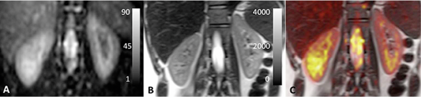
On the molecular level healthy and diseased tissues show very different diffusion characteristics.
Prof. Siegfried Trattnig
A very straightforward example of an imaging biomarker is the size and volume of a tumour determined in computed tomography. But spectroscopy and nuclear medicine also apply quantifiable parameters. In tumours, for example, changes can occur in the cell membrane involving the metabolite choline: an increased choline concentration in tissue detected by spectroscopy indicates a malignant tumour. In nuclear medicine tracers that dock onto particular metabolites are injected into the body.
In magnetic resonance imaging (MRI) – Trattnig’s specialty, T1 and T2 relaxation times and the apparent diffusion coefficient (ADC) are measured. ‘On the molecular level healthy and diseased tissues show very different diffusion characteristics,’ Trattnig explained. The more cells in the tissue, the higher the water diffusion and ‘since a tumour consists of many more cells than healthy tissue it shows different diffusion characteristics and thus pathological changes can be detected much earlier than in a merely morphological analysis’.
In cell diseases the ion pump becomes impaired
The Trattnig team at Vienna’s High-Field MR Centre works on sodium MR imaging, which is based on the fact that the sodium-potassium pump in healthy cells ensures that the sodium concentration within cells is higher than without. When a cell diseases, the ion pump is one of the first elements of the cell to be impaired. Consequently, tumour cells show a much higher sodium concentration than healthy cells.
The European Imaging Biomarker Alliance
This new MRI approach measures intracellular and extracellular sodium levels and ‘allows us to detect and quantify the earliest stages of cancer on the cellular level, the professor said. This might be relevant, for example, in breast cancer therapy: current imaging technologies can visualise tumour response to chemotherapy after four to six weeks; sodium MRI might be able to do that within days.
To support these new developments, last year the European Society of Radiology (ESR) re-organised its imaging biomarker activities: several subcommittees and working groups on imaging biomarkers were combined to form a single unit, the European Imaging Biomarker Alliance (EIBALL).
‘We aim to move imaging biomarkers to clinical application,’ explained Trattnig, the EIBALL chairman.
EIBALL cooperates with other organisations, such as the Quantitative Imaging Biomarker Alliance (QIBA), which has been coordinating the biomarker imaging activities in the USA for several years. ‘QIBA drove technological development and designed a number of standardised parameters,’ Trattnig said. ‘However, large multicentre studies to compare several parameters cannot be conducted in the USA for bureaucratic reasons. In Europe it’s much easier to apply biomarkers in large clinical studies.’
EIBALL also works closely with the European Organisation for Research and Treatment of Cancer (EORTC), the coordinator of all large European multicentre cancer studies. ‘Until recently, oncological societies disregarded imaging due to the lack of standardisation; but now radiologists are members of the disease-oriented groups of EORTC,’ Trattnig is happy to report. Thus, for the first time, radiologists are involved in the design of such studies and can ensure that imaging biomarkers are included.
Profile:
Dr Siegfried Trattnig is a Professor of Radiology with a focus on high-field MRI at the Medical University in Vienna, Austria. He has been Medical Director of the high-field MRI research scanner since 2000 and, from its founding in 2003, of the High-Field MR Centre (HFMRC) at MedU Vienna. He is also a member of more than 50 scientific committees in all major international radiology, orthopaedics and MRI societies, and has chaired the European Imaging Biomarker Alliance (EIBALL) since its establishment in 2015.
03.03.2016