Source: Photonics21
News • Photonics21
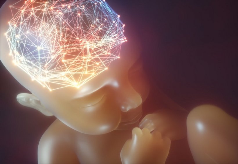
Infrared scanner spots brain damage in babies early
An estimated 500,000 babies born around the world each year develop unnecessary brain damage that could be treated if caught in time – but monitoring these infants' delicate brains is extremely difficult. A new photonics device currently in development aims to reduce unnecessary disabilities by improving the instant, real-time monitoring of newborn babies with harmless light particles.
No medical tools currently exist to create a harmless, real-time, continuously moving image inside newborn babies' delicate brains. MRI scans can provide an accurate picture inside adults but are highly unsuitable for newborn babies, given they require a patient to sit still while giving out harmful radiation.
Neurodevelopment disabilities like cognition or motor skill impairments that affect half a million infants globally every year – resulting from defective heart vessels – can be treated but are difficult to monitor and catch in time. However, the ‘TinyBrains’ health consortium run in conjunction with ICFO - The Institute of Photonic Sciences in Barcelona is developing a new wearable device to help doctors see what is going on inside infants’ minds quicker than ever.
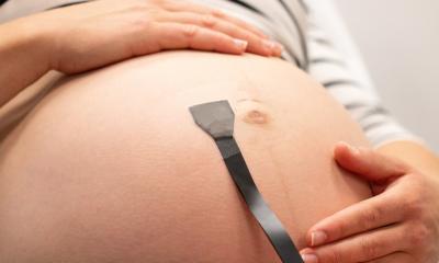
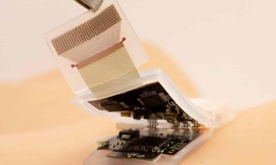
Putting near-infrared lasers and LEDs into a small, wearable cap that are combined with EEG electrodes, the scientists send harmless signals into the infant’s brain – working almost like an ultrasound scan, but using photonics (or light) to give much more information, a more detailed picture and an image of the underlying brain activity rather than the anatomy.
The signals can measure the cause of so many unnecessary neurodevelopment disabilities by keeping a close eye on any slight drops in critical oxygen levels to and from the brain instantaneously in real-time.
Heart defects and neurological complications
TinyBrains project coordinator, Professor Turgut Durduran, said: "A staggering 500,000 people suffer unnecessary disabilities that result from congenital heart defects (CHD) and other structural defects in the heart across the world, drastically affecting the life of the patient if they are not picked up soon after birth. At present, it is tough to monitor these at-risk populations both technically, because of the lack of appropriate tools, and also ethically because consent and risks have to be taken into consideration."
Each year 3.4 million babies worldwide are born with a congenital disability, and of these, congenital heart defects (CHD) are the most frequent. About 40% of these infants need a cardiac surgical intervention during their first year of life with a subsequent stay in the intensive care unit.
Most of these babies survive to adulthood but risk suffering from deficits in their neurological development due to brain blood flow and perfusion alterations happening during the intervention. These alterations often result in learning disabilities, leading to low quality of life for these patients and their families, constituting a significant challenge to public health.
Scanning with light
The cap's sensors connect to a portable unit and measure the cerebral metabolic rate of oxygen – or the oxygen saturation in the blood and the concentrations of oxy- and deoxy-haemoglobin – and build up a 3D colour image in real-time. “We are using high-density near-infrared spectroscopy (fNIRS) and diffuse correlation spectroscopy (DCS) to measure the oxygen saturation levels in the blood. By integrating both of them with an imaging device as the electroencephalography (EEG), the resulting 3D images have higher resolution, increase the brain specificity and penetration and for the first time, a spatial resolution to this class of measurements.”
By identifying brain function alterations during surgery and stays in intensive care units will allow doctors to analyse why brain disorders frequently occur in the postnatal period and to pinpoint the types of clinical interventions that can improve the neurological outcome of these infants and, ultimately, their quality of life, as infants, young persons and adults.
Source: Photonics21
28.04.2021