Radiation treatment decisions in patients with prostate cancer and suspected lymph node metastasis based on USPIO-MRI
For the second in his series of articles for European Hospital, Professor Stefan Schönberg of the Institute of Clinical Radiology and Nuclear Medicine (IKRN), University Hospital Mannheim, Medical Faculty Mannheim, University of Heidelberg, invited colleagues from the Nijmegen University Medical Centre (UMC St Radboud) for a roundtable discussion on:
Prostate cancer is the most frequent cancer in men with about 49,000 newly diagnosed cancers in Germany and 6,000 in the Netherlands, annually.

Important treatment options for local prostate cancer are radical prostatectomy (RPx) and radiation therapy (RT). Currently, after initially performed transrectal ultrasound, multimodality high resolution MR imaging is the preferred method for accurately assessing the local disease stage and is performed to guide decision making for optimal treatment choice. Multimodality MR imaging includes multiplanar T2-weighted images to show morphologic attributes, chemical shift spectroscopy with further mapping of the Choline/Citrate ratios for measurement of alteration in metabolism, dynamic sequences for delineation of typical cancer hypervascularisation and diffusion-weighted sequences to show restriction in the Brownian movement of the water molecules. Image quality and spatial resolution are improved by utilising a balloon type endorectal coil. In an innovative procedure, an endorectal balloon is also used for CT imaging and during radiotherapy. Thereby, the prostate will have similar shape and the rectal tissue will be protected from radiation damage because it is displaced away.
Another important field in prostate imaging is lymph node staging. This comprises measurement of pelvic lymph node sizes by CT or MRI and stratifying patients in risk groups for possible lymph node metastasis according to the Partin or Narayan tables. These depend on Gleason-Score (histological grading) and PSA value. However, conventional CT and MRI can miss metastases in small, non-enlarged lymph nodes. Furthermore, in patients at low risk for nodal metastases based on these tables, no additional nodal diagnostic imaging is performed.
Recently, diagnostic imaging techniques have become available which enable improved lymph node staging. At present, the most accurate method is MR lymphography with ultra-small super paramagnetic iron oxide particles (USPIO, Sinerem). Using a 3T scanner, metastases in nodes measuring 2-3 mm can be detected. Up to now no serious side-effects have been reported. Regrettably, this promising technique, using Sinerem contrast, is not yet commercially available.
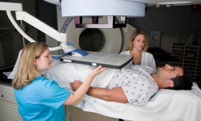
Currently, the extension of radiation fields depends on the risk for lymph node metastasis. This risk is calculated according to the Roach formula. Patients at low risk (<15%) undergo irradiation of the prostate only, whereas patients with high risk (>15%) may benefit from radiation of the whole pelvis, including the lymph nodes. Moreover, it has been demonstrated that radiation therapy is more effective at a higher dose. This requires an exact delineation of both tumour and metastases. Following this, improved radiation techniques, like intensity-modulated radiation therapy (IMRT), are needed for patients to benefit from the advanced imaging. In a new concept, the ‘dominant intraprostatic lesion’ (DIL) is irradiated with a higher dose than the rest of the prostate. A similar procedure is planned for the concept of ‘MR guided lymph node IMRT’ (MGL-IMRT).
Currently, the above-mentioned multimodality MR imaging, as well as the USPIO-MRI for the lymph nodes, is only available at specialised centres and universities. To further assess the value of these techniques, for improvement and routine implementation, a collaboration has been set up between the Departments of Radiology and Radiation Oncology in both, the University Medical Centre St. Radboud, in Nijmegen, and the University Hospital in Mannheim. These two centres will closely work together on MR-imaging of the prostate to perform bi-centre trials and to facilitate further multicentre trials.
In this way the potential of improved prostate imaging to enable a more effective therapy will be optimally explored. As a consequence of this, it is envisioned that disease-free survival of patients with prostate cancer can be prolonged and side-effects and costs caused by not stage adapted treatment can be reduced.
Additionally, we would like to invite you to the newly established transatlantic meeting ACSI, which will take place on 20-21 June 2008 in Mannheim, Germany.
Details: www.mr-pet-ct.com
30.04.2008


