The crucial role of radiology in prostate cancer diagnosis and therapy
Urologist Arnulf Stenzl: "Imaging is the key to prostate cancer treatment"
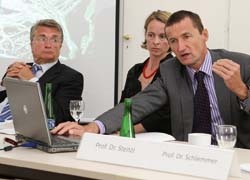
Emphasising the crucial partnership of radiologists and urologists in the treatment of prostate cancer, Professor Arnulf Stenzl, Medical Director of Urology at the Tübingen University Hospital, explained that, throughout the phases of prostate cancer diagnosis and therapy - from primary diagnosis onward - imaging is indispensable.
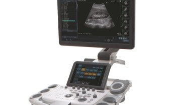
Elastography, an ultrasound imaging technique that is increasingly the procedural choice for the initial detection of prostate cancer because it can precisely locate a tumour in the chestnut-size prostate gland, assesses tissue elasticity – an important indicator because cancer tissue is often denser and firmer than healthy tissue. ‘For the urologist, elastography is thus, in a way, a computer-assisted further development of rectal palpation,’ Prof Stenzl explained. Exact localisation increases the success rate of biopsies: ‘With biopsies we always run the risk of sampling healthy rather than tumour tissue. Elastography can help reduce the rate of false-negative results.’
If the biopsy provided a positive prostate cancer result, the next step is to determine whether the tumour is confined to the prostate or has spread to other tissue or organs. For this, MRI is preferred, which is also an important surgery planning tool, because MR images can show the amount of tissue that needs to be removed.
An interesting and very promising method is magnetic resonance spectroscopy (MRS), which provides very good spatial resolution of the organ, as well as insights into the chemical composition of the tissue. A healthy prostate produces citrate, which inhibits urolith formation in the body. Cancerous prostate tissue shows low levels of citrate and high levels of choline, a cell building block needed for the construction of cell membrane. Increased choline levels indicate tumour growth. Low citrate and high choline levels in the prostate can be detected easily with spectroscopy, which analyses different resonance frequencies.
Prostate cancer frequently spreads to other body areas, particularly the lymph nodes and bones. Choline PET-CT, a system that combines diagnostic imaging and the visualization metabolic events, is used to detect metastases.
‘Such combined modalities allow the detection and exact localisation of minute tumours, thanks to the slice images provided by computed tomography,’ Prof Stenzl pointed out.
In addition, the tumour marker choline comes into play: as C11-choline, combined with radioactive carbon, the substance is intravenously injected into the body. PET/CT detectors catch signals emitted by the radioactive tumour marker for conversion into an image. ‘The intensity of the signals indicates where in the body’s metabolism choline is particularly active. We can thus localise cell growth caused by prostate cancer. Currently, this is the most sophisticated diagnostic method, but it needs further improvement,’ he said.
In the long term, Prof Stenzl predicts that super-precise MRI will replace computed tomography: ‘That’s the direction we urologists would like radiology to take. But the fact that we voice certain wishes will in no way belittle the enormous support we currently receive from imaging diagnostics.’
01.09.2009


