Source: Adobe Stock/phonlamaiphoto
News • MetaboliQs
Hyperpolarized nuclear MR: more precise diagnoses
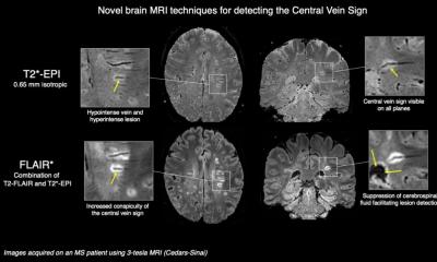
Hyperpolarized nuclear magnetic resonance enables major medical advances in molecular diagnostics, for example for cardiovascular diseases or cancer therapy. Within the framework of the EU collaborative project “MetaboliQs”, seven partners developed a microscopy method which enables the analysis of metabolic processes at the single cell level by means of diamond-based hyperpolarization. In addition, the consortium successfully demonstrated hyperpolarization using the PHIP method in application-oriented MRI experiments.
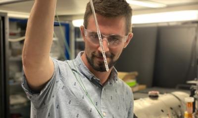
Coordinated by the Fraunhofer Institute for Applied Solid State Physics IAF and NVision Imaging Technologies GmbH, an international consortium of seven research institutions and industrial companies has achieved breakthroughs in quantum microscopy for the analysis of metabolic processes and the application of Parahydrogen Induced Polarization (PHIP) within the project “MetaboliQs—Leveraging unparalleled room temperature quantum coherence to enable safe, first-of-its-kind, multimodal cardiac imaging”. The results significantly advance two promising approaches for improving medical imaging diagnostics and spectroscopy by making use of nuclear magnetic resonance (NMR) more precise, practical and efficient. Under the Future and Emerging Technologies (FET) program “The Quantum Flagship”, the European Union (EU) funded “MetaboliQs” since 2018.
On the one hand, the collaborators exploited the special quantum sensing properties of nitrogen-vacancy centers (NV centers) in nanostructured diamond to detect NMR signals with 1000-fold better spatial resolution compared to the current state of the art, proving that microscopic spectroscopy is suitable for metabolic analyses on single cells. On the other hand, the researchers successfully demonstrated for the first time that a PHIP quantum polarizer is feasible for high sensitivity preclinical in vivo studies, demonstrating hyperpolarized magnetic resonance imaging (MRI) under real-world conditions.
Dr. Volker Cimalla, project leader at Fraunhofer IAF, classifies the results of the project: “Our approach aimed at bringing the unique advantages of diamond-based quantum sensing to medical applications. With the developed quantum microscope, we have created a unique research tool that decisively advances cell analysis and opens up new possibilities for medical research and in vitro diagnostics.” Ilai Schwartz, project coordinator on the part of NVision, emphasizes: “The developed quantum polarizer paves the way for a promising technology to realize hyperpolarized MRI. Compared to current methods, the PHIP approach has the advantage of being significantly faster, more practical, and more resource-efficient while maintaining maximum precision.”
Recommended article

Article • Imaging tumour metabolism
Hyperpolarised MRI boosts cancer diagnosis
Tumour metabolism can be imaged with MRI as a technique to help determine cancer aggressiveness and response to therapy. The work by a UK-based group, on probing cancer metabolism non-invasively with clinical hyperpolarised carbon-13 MRI, can detect metabolic changes in the tumour. As metabolic changes occur much earlier than change in tumour size, this could have implications for quicker…
Diamond-based hyperpolarization improves NMR
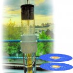
Hyperpolarization can overcome the major drawback of NMR technology: its relatively low sensitivity. Classically, NMR spectrometers or MRI systems measure the electrical signals generated when nuclear spins within an external magnetic field respond to a resonant radio frequency pulse. The signal strength here depends on the thermal polarization of the sample under study, i.e., the number of nuclear spins magnetically aligned within it. The signal is usually very weak, since on average only one out of several billion nuclear spins is magnetically aligned. However, hyperpolarizing techniques magnetically align a large fraction of the nuclear spins for a certain period of time, which increases the strength of the NMR signal by several orders of magnitude.
Therefore, with an NMR signal amplified 100,000-fold by hyperpolarization, medical applications such as MRI can be improved many times over. Cardiovascular diseases could can be diagnosed much earlier, cancer therapies could be immediately tested for their effect and thus be personalized, since physicians are able to detect typical metabolic processes at the molecular level in real time. For this reason, researchers worldwide are working on various approaches to develop viable hyperpolarization methods for medical applications. Current methods such as dynamic nuclear polarization (DNP) are already very precise, but extremely resource-consuming. Moreover, the hyperpolarized state lasts only for seconds.
Recommended article
News • Singlet contrast imaging
Hyperpolarized MRI enables real-time view on metabolic processes
Hyperpolarized MRI is a recent development and its research and application potential has yet to be fully explored. Researchers at Johannes Gutenberg University Mainz (JGU) and the Helmholtz Institute Mainz (HIM) have now unveiled a new technique for observing metabolic processes in the body. Their singlet-contrast MRI method employs easily-produced parahydrogen to track biochemical processes in…
From material growth to medical research
In this light, the collaborative partners of the “MetaboliQs” project relied on the special quantum physical properties of NV centers in synthetic diamond, which Fraunhofer IAF grew and nanostructured on the optimized material provided by Element Six (E6). The Hebrew University of Jerusalem (HUJI) analyzed the quantum properties in the structures. NVision realized the prototype quantum microscope based on the characterized nano diamond chips and demonstrated in feasibility studies both hyperpolarization using optically polarized electron spins in diamond and detection of hyperpolarized metabolites at high spectral resolution. Bruker BioSpin GmbH was responsible for evaluating the samples from NVision to determine spin concentration and relaxation time. With the setup, the collaborative demonstrated NMR spectroscopy on metabolites on a microscopic scale for the first time.
Finally, in preclinical in vivo comparison studies, the Technical University of Munich (TUM) showed that the quantum polarizer also provided by NVision, which enables hyperpolarization by transferring parahydrogen to 13C nuclei, is superior to methods based on dynamic nuclear polarization: with a fraction of the resources required, the degree of polarization and concentration as well as the relaxation time of metabolic tracers are significantly higher than with the alternative method. Researchers at the Swiss Federal Institute of Technology Zurich (ETH Zurich) successfully simulated that a better signal-to-noise ratio in imaging can be achieved if the strength of the magnetic field is reduced from 3 T to 1.5 T or 0.75 T. Thanks to the superior properties of the PHIP quantum polarizer, MRI results are qualitatively on par even with a weaker magnetic field; in return, the cost of operating the MRI system drops significantly.
Source: Fraunhofer Institute for Applied Solid State Physics IAF
23.05.2022