ECR 2015
Challenges slow down integration of imaging biomarkers in Europe
Integration of imaging biomarkers in Europe is a challenge, and a number of issues must be overcome so that the full potential of quantitative imaging can be realised, renowned researchers will show in a session today at the ECR.
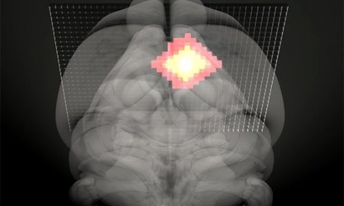
Quantitative imaging enables disease to be found very early and considerably improves treatment monitoring. In healthy tissue for instance, researchers can use quantitative imaging to study the functional properties of this tissue in order to get the most important meaning for biologists, physicists, etc., in a non-invasive way. Based on this information, they can develop biomarkers, which help to study the disease and its history, use the data to predict outcome and, most importantly, to determine the responsiveness of individual person to therapy – and assess its efficiency.
Applications for imaging biomarkers include the detection and treatment of cardiovascular diseases, neurological diseases, musculoskeletal disorders, metabolic diseases, autoimmune pathologies and inflammation. Biomarkers also play a key role in the development of new drugs.
Several bottlenecks currently prevent quantitative imaging from fully benefiting healthcare. Imaging biomarkers have to be technically validated, robust and reproducible; then they must be clinically validated. This process is extremely long. To make matters worse, imaging biomarkers are not included in the landscape of European biobanks.
“There is a great and urgent need to include biomarkers in European biobanks, since imaging repositories are dealing with big data, and have specific technical requirements in terms of codification, standards and interoperability,” said Prof. Guy Frija, consultant radiologist at the Imaging Department at Hôpital Européen Georges Pompidou in Paris, who will chair the session.
Frija, who is Past-President of the European Society of Radiology, pushed for the society’s establishment of a European Biomarkers Task Force, creating synergies with RSNA’s Quantitative Imaging Biomarkers Alliance and tackling clinical validation of biomarkers at a European level.
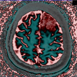
Quantitative imaging must also overcome a number of technical difficulties. MR offers a variety of techniques – diffusion imaging, dynamic contrast enhanced MR and spectroscopy – to measure tissue and obtain biochemical information on the tissue of interest. But MR examinations are not reproducible and this is a major obstacle for quantification, according to Prof. Siegfried Trattnig, Professor and Head of the Centre of Excellence for High Field MR, Department of Biomedical Imaging and Image-guided Therapy at Vienna General Hospital, who will also speak during the session.
In addition, performing quantitative imaging takes time compared with regular scans. Patients must lie still for longer in the scanner for multiparametric data to be obtained, so they are more likely to move and this can hamper data quality. “What we really need is a fast technique and the best would be if we could acquire multiple parameters simultaneously,” he said.
This may change with recently developed sequences, in which researchers can acquire T1 and T2 relaxation times of a certain tissue or region with the same sequence. But the biggest advance may yet come from MR Fingerprinting (MRF), which completely changes data acquisition, post-processing and visualisation. Trattnig will present these advances in detail during his talk.
Source. Press release European Society of Radiology
05.03.2015