Cardiac ergometry in an MRI
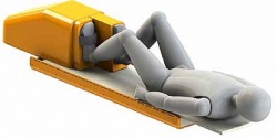
Ergospect develops ergometers for muscle exercises inside the magnetic resonance bore. The company has added two new MRI-compatible ergometers to existing devices for musculoskeletal examinations. The Diagnostic Pedal Cardio, for cardiac stress MRIs, enables examination of perfusion, motility and energy metabolism of the myocardium under stress. Today, MRI magnetic resonance imaging of the heart during physical stress is carried out routinely. Due to the lack of suitable devices, drugs such as Dobutamin or Adenosin are usually used to stress the cardiovascular system. However, this does not reflect the physiological reality. The Diagnostic Pedal Cardio modules are suitable for MR and have been specially designed to stress the heart in an MRI bore. Thus it is possible to investigate the performance and perfusion of the myocardium during a dynamic exercise via MRI or magnetic resonance spectroscopy (MRS), which provides an effective early diagnosis of risk patients and a more accurate diagnosis of a patient with coronary heart disease (CHD) and with myocardial infarction (MI). All pedals are compatible with all MRI-systems (up to 7-Tesla) and consist of a basic platform to be combined with different modules. The firm will also develop and deliver individual solutions according to customer specifications.
Details: www.ergospect.com
02.11.2012