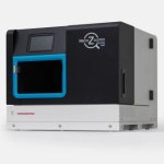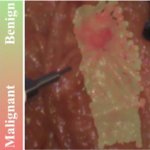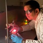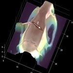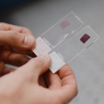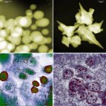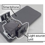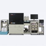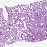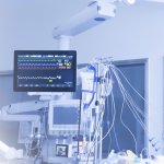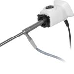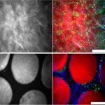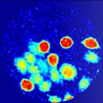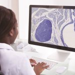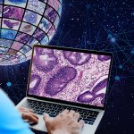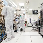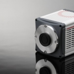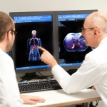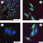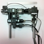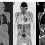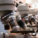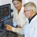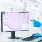
News • From slide preparation to imaging
New global partnership addresses digital pathology workflow
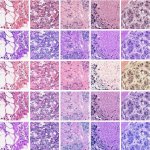
Sakura Finetek and Hamamatsu Photonics K.K. have announced a collaboration aimed at standardising slide preparation and digitisation to support diagnostic accuracy in pathology.