First hologram video player to show your beating heart
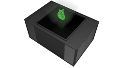
UK scientists are developing an interactive holographic video created from an MRI or CT scan that can display live footage of internal organs in front of a user where features can be rotated, enlarged, and isolated, delivering a breakthrough in medical imaging and education.


Popping in to your local hospital may be much more revealing in as little as three years thanks to engineers at Holoxica Limited, who have invented a moving 3D video hologram. Watching your heart beat, your lungs inflate or your unborn child in life size and before your eyes as a hologram that can be rotated or enlarged, in real time is no longer the stuff of science fiction.
With no need for 3D specs or a virtual reality headset, the dynamic or ‘moving video’ 3rd Generation holograms are made by gathering multiple ‘slices’ of an internal organ, such as a brain or a liver, from a normal CT or MRI scanner. These ‘slices’ of data are then assembled through a ‘diffractive holographic screen’, producing single colour green pixels, or ‘voxels’, in mid-air and essentially bending light to the will of the user.
Teaming up the European photonics innovator accelerator ACTPHAST, hologram specialists Holoxica have linked photonics technology with their 1st and 2nd Generation holographic motion displays to develop one of the most revered gadgets of science fiction, an idea that never seemed to take off in real life. Holoxica’s CEO, Dr Javid Khan explains: “Hollywood depicts holographic displays as something ubiquitous in films from Iron Man to Avatar. This has created inflated expectations in the mind of the public who largely believe that displays or ‘holographic projectors’ already exist and are trivial to make. This is not the case.”
Instead of trying to create a mythical “Star Wars” display, Holoxica took a more pragmatic approach by starting with the simplest holographic display, a single pixel, or ‘voxel’, in 3D space, that could be switched on or off.
“After the first voxel, we moved on to two, working up to 4, then 9, then 16 voxels and so on. Our images are not projected; they are holographically reconstructed using diffractive optics. Projection implies scattering off a surface, but here there is no surface, only air. We are using photonics design and engineering of diffractive optical elements to bend or form light to produce images in mid-air. Although we are looking at targeting medical, scientific and engineering imaging fields to start with, holographic video will change gaming, communication and create a new digital revolution,” Dr Khan enthused.
With the possibility to isolate features, zoom in, rotate and pan around 3D space, the 3rd Generation dynamic display presents an array of exciting opportunities for the future of surgery and anatomical study. “Take current imaging techniques like CT scans where radiologists are trained to interpret the multiple levels of data, or ‘slices’ of the brain. Medical consultants, specialists and surgeons are not trained to do this and therefore need to build up a mental stack of the scans or rely on second-hand interpretation.”
“For the first time, a physician will be able to see a tumour in an impossible part of the brain and make an informed decision. This is also easier for patients to understand what is going on. Teaching anatomy with this device will give students a hitherto unrivalled understanding.”
Source: Holoxica Limited
03.01.2017


