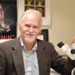
News • Importance of tumor location
Testosterone slows glioblastoma growth in men
New research reveals that testosterone slows glioblastoma growth—the opposite of the androgen’s effect on tumors outside the brain.

New research reveals that testosterone slows glioblastoma growth—the opposite of the androgen’s effect on tumors outside the brain.

Researchers are developing nanozymes to improve treatment of aggressive brain tumours. The tiny particles can be activated by near-infrared light and applied directly during surgery.

Researchers have developed a new MRI-based method that enables objective quantification of the growth of the most aggressive brain tumours, particularly glioblastoma.

Patient-derived organoids (PDOs), or tumoroids, for pediatric brain cancer show promise in helping researchers find new drugs and better understand the different responses of the tumor.

Researchers discovered that pineoblastoma, retinoblastoma and medulloblastoma – severe brain tumours in children that appear to be completely different – actually arise from the same type of cell.

Functional brain radiosurgery is an application of stereotactic radiosurgery (SRS), representing its newest clinical field. It is a precise, non-invasive medical technique using focused ionising radiation to precisely target specific brain structures to modulate brain function for neurological disorders, psychiatric conditions, or intractable pain. The technology offers “precision without…

Clues in the CSF: Researchers have developed the first high-precision method that can theoretically diagnose common brain tumors in children and adolescents without surgery.

A new kind of intraoperative MRI (iMRI) can perform functional MRI scans in real time during brain surgery — helping surgeons detect potential complications in as little as seven seconds.

To soften up tumors prior to cancer chemotherapy, a team of researchers paired high-frequency ultrasound waves with a type of sound-responsive particle to reduce the protein content of the tumors.

A novel AI-based method can distinguish between progressive brain tumours and radiotherapy-induced necrosis on advanced MRI. This could help clinicians more accurately identify and treat the issues.

IDH mutated gliomas are slow-growing brain tumors with a relatively good prognosis. A new study shows that many patients reveal measurable cognitive impairment in the first year after treatment.

Delivering a pathology service in resource-constrained locations and developing countries remains a challenge. Cost is a significant barrier, as is the availability of equipment, trained staff and technical and IT support can also hinder a desire from clinicians and pathologists to give their patients a high level of service to help their diagnosis and recovery. The subject was tackled in a…

Philips and Cortechs.ai have extended their collaboration to integrate AI-enabled quantitative neuroimaging analytics directly into MR systems. The partnership aims to provide radiologists with automated, objective measurements of brain structures and lesions to support diagnosis and monitoring of neurological conditions.

NaoTrac is a fully autonomous surgical navigation robot designed to enhance precision, safety, and efficiency in neurosurgery. By integrating advanced technologies with the expertise of surgeons, NaoTrac streamlines surgical workflows, shortens the learning curve, and improves patient outcomes.

Researchers identified a targeted way to protect the brain from harmful side effects of cranial radiation therapy, potentially preserving the quality of life for millions of brain cancer survivors.

At the EANS neurosurgery congress in Vienna, Italian medical imaging company Esaote presented I-Genius, a new open MRI system designed to provide real time checks during glioma surgery.

Scientists have shown for the first time that glioblastoma—the deadliest form of brain cancer—affects not just the brain but also erodes the skull, alters the makeup of skull marrow, and interferes with the body’s immune response.

Melanoma testing could one day be done at home with a skin patch and test strip with two lines, similar to Covid-19 home tests, according to University of Michigan researchers.

A new method to monitor blood flow in the brain helps neurosurgeons detect the risk of a stroke during surgery – and potentially prevent it. This could also be useful for other types of operations.

A new AI-based tool measures cancer aggressiveness by analyzing the ‘stemness’ of tumors – their similarity to pluripotent stem cells. This could pave the way for new therapies.

Using a keyhole surgery approach, surgeons have found a new way to access previously difficult-to-reach brain regions with faster recovery times. With the help of 3D modelling technology, the neurosurgeons succeeded in removing complex tumours from the cavernous sinus through the eye socket, avoiding complex brain surgery and enabling their patients to make a quicker recovery.

Childhood cancer diagnosis times span from immediate to delays of several years, according to a new study. Young bone tumor patients are among the most affected by these delays.

As Emergency Departments (EDs) get ever busier, focus has fallen on the role artificial Intelligence (AI) can play in supporting patients and clinicians in delivering urgent care. The topic took centre stage in a session looking at the ethics of AI in the ED at the annual congress of the European Society of Emergency Medicine (EUSEM) in Copenhagen.

New research reveals that self-generated electrical activity of small cell lung cancer (SCLC) directly promotes tumor metastasis and progression. This may provide new avenues for future treatments.

Using a new technology and computational method, researchers have uncovered a biomarker capable of accurately predicting the aggressiveness of meningioma brain tumors and breast cancers.

A new blood test that detects a specific brain protein may help doctors determine the type of a stroke faster and allow them to start safe treatment for people before they get to the hospital.

Researchers have developed an artificial intelligence (AI) model to detect the spread of metastatic brain cancer using MRI scans, offering insights into patients’ cancer without aggressive surgery.

Precise segmentation of anatomical structures greatly benefits cancer diagnosis. Using AI and deep learning methods, researchers are developing a high-precision 3D viewer software for medical image data.

Monitoring brainwaves and diagnosing neurological conditions could benefit from a novel 3D printing technology, which applies liquid ink onto a patient’s scalp to measure brain activity.

US researchers have developed a comprehensive deep learning AI model designed to more accurately identify and classify cells in high-content tissue images.

A new approach to fight glioblastoma: Swiss researchers have now developed an immunotherapy that not only attacks the brain tumor—it also turns its microenvironment against it.

In many cases, metastatic breast cancer is still incurable. To improve the odds, researchers have now analyzed the diversity of metastatic cells and their interactions with their cellular environment.

Researchers have identified fibrotic scarring as a key source of resurgence of glioblastoma multiforme (GBM). The new insights could lead to better prevention of this type of brain cancer.

To diagnose neuroblastoma in children, lengthy scans and anaesthesia are often required. A new PET/CT imaging technique could deliver faster results without sedation for the paediatric patients.

Researchers discovered islands of highly potent immune cells in the vicinity of glioblastomas. This may open up prospects for new therapies for these aggressive brain tumours.

The huge amount of data generated by modern MRI scanners presents challenges in storage, accessibility, and security. To that end, Chinese researchers have developed a cloud MRI system.

Neurosurgeons have developed a minimally invasive surgical technique to remove lesions in the petrous apex. The method provides a new approach to these notoriously diffcult-to-remove tumors.

A new prototype self-powered sensor could improve the process of medical imaging for patients and technicians by detecting movement and shutting down an MRI scan in real time.

A new study used new molecular analyses to unravel the biological mechanisms of pediatric brain tumors and refine their classification.

Artificial cells to combat cancer: Research groups are working to create synthetic micro-organisms capable of detecting the presence of the disease and delivering anti-cancer therapies.

A new sensor prototype can detect errors in MRI scans in a way that is impossible for current electrical sensors – and hopefully pave the way for MRI scans that are better, cheaper and faster.

Diamond dust could be used in MR imaging, a surprising find shows. The nanometer-sized particles could serve as an alternative to the controversial contrast agent gadolinium.

A study highlights performance differences in computational pathology systems, depending on demographic profiles associated with histology images. The researchers also propose a way to fix this bias.

A novel AI-based, non-invasive diagnostic tool enables accurate brain tumor diagnosis, outperforming current classification methods. The tool leverages MRI information to aid clinical decision making.

Holding a mobile phone close to the head for an extended amount of time has long been connected to brain cancer. Now, a new study found no hints for an increased risk.

Targeting a specific protein, Finnish researchers may have found a way to restore drug-resistant neuroendocrine prostate cancer to a state that responds to treatment.

Breast surgery is a traumatic experience for a woman, no matter whether breast-conservation surgery (BCS) or a mastectomy. Trauma levels are greatly enhanced, if pathological evaluation findings of an excised breast tumour following a lumpectomy suggest that additional cancer may still be in the margins, and a second surgical procedure is required. A new system with the ability to accurately…

White blood cells found in breast tumors can both help and hinder the spread of cancer cells to other organs, a new study from Karolinska Institutet shows.

Research from Shenzhen proposes an integrated diagnosis model for automatic classification of adult-type diffuse gliomas directly from annotation-free standard whole-slide pathological images.

Machine learning and AI are playing an increasingly important role in medicine and healthcare, and not just since ChatGPT. This is especially true in data-intensive specialties such as radiology, pathology or intensive care. The quality of diagnostics and decision-making via AI, however, does not only depend on a sophisticated algorithm but – crucially – on the quality of the training data.

Advances in positron computed tomography (PET) could lead to a more refined approach to the precise removal of brain tumors is on the horizon, experts from Poland point out.

A new type of microdevice could offer new ways to treat brain cancer. The shape and size of a grain of rice, it is implanted into a tumor to study the effects of ongoing therapies.

Researchers at NIST have been working on several fronts to advance low-field MRI technology and validate methods for creating images with weaker magnetic fields.

Scientists have designed an AI tool that can rapidly decode a brain tumor’s DNA to determine its molecular identity during surgery — critical information that can guide treatment decisions.

A new US-led study shows that astrocyte brain cells play an important role in promoting brain metastasis by recruiting a specific subpopulation of immune cells.

US researchers identified a potential breakthrough in glioblastoma treatment. Using a modified virus, they created a treatment that specifically attacks tumor cells, while leaving normal cells intact.

Damage to the brains of patients operated on for brain tumors may be assessed by measuring biomarkers in the blood pre- and postoperatively, a new study from the University of Gothenburg shows.

For decades, researchers have marveled at the ability of glioblastoma, a particularly aggressive brain cancer, to turn off a patient's cancer-fighting immune cells, thereby allowing tumors to grow freely.

US researchers have developed a way to use MRI scanning to map body cell metabolism, opening up new possibilities for detecting cancers and revealing if a tumor is responding to treatment.

A novel imaging modality that can visualize the distribution of medical radiopharmaceuticals with very fine resolution has been developed and successfully tested.

A new real-time imaging technique that uses a type of infrared light has, for the first time, been used during surgery to differentiate between cancerous tumours and healthy tissue.

The world's first MRI scanner with a magnetic field strength of 14 Tesla will be built in Nijmegen. Its high sensitivity will allow scientists to image the brain in more detail.

Philips is an active participant in the new European Federation for Cancer Images consortium (EUCAIM) – a project launched in the run-up to World Cancer Day 2023.

A Korean research team successfully changed the properties of carcinogenic cells in the lungs and eliminate both drug resistance and their ability to proliferate out to other areas of the body.

It was previously assumed that bones lacked lymphatic vessels.Now, new research not only locates them within bone tissue, but demonstrates their role in bone and blood cell regeneration.

The distinction between primary tumors and metastases can be made quickly and accurately in brain tumors using radiomics and deep learning algorithms, a new study shows.

Engineers have developed an electronic patch that can give medical professionals unprecedented access to crucial information that could help spot life-threatening conditions.

Glioblastomas march to the beat of a different drum: researchers discovered a type of cells that act as pacemakers, driving the growth of the brain tumour and increasing its resistance.

For the first time, a new study has identified enlarged perivascular spaces in the brains of migraine sufferers. Results of the study will be presented at the annual RSNA meeting.

Using a special type of MRI, researchers have uncovered brain changes in patients up to six months after they recovered from Covid-19, according to a study being presented at the annual RSNA meeting.

Treatment of central nervous system diseases and tumors is often hindered by the blood-brain barrier. A new method aims to overcome this obstacle using focused ultrasound intranasal delivery (FUSIN).

AI-based diagnosis is undoubtedly one of the most promising subjects when we talk about the future of radiology. Now, a couple of new studies indicate that most radiologists are open to using the technology and this for good reasons.

The classification of brain tumors—and thus the choice of optimal treatment options—can become more accurate and precise through the use of artificial intelligence in combination with physiological imaging.

Reseachers have developed a novel microneedle for injecting therapeutics into the eyes, potentially solving one of the major challenges of treating eye diseases.

A research team in Spain and the US has created 3D-printed acoustic holograms to improve the treatment of diseases like Alzheimer's and Parkinson's, among others.

A protein vital in determining the organs affected by metastasis has been identified by Swiss researchers. This could lead to the development of therapeutic approaches to suppress metastasis.

MediSCAPE, a high-speed 3D microscope, can see real-time cellular detail in living tissues to guide surgery, speed up tissue analyses, and improve treatments.

For a long time, the origin of metastasis remained obscure. Now, scientists have discovered some of the mechanisms these cells arise.

Children's National Hospital successfully performed the first-ever high-intensity focused ultrasound surgery on a pediatric patient with neurofibromatosis. This is the youngest patient to undergo HIFU treatment in the world.

Researchers at the University of Sussex are one step further to developing a blood test capable of diagnosing the most aggressive form of brain tumour.

In both the mice and organoids, cytokines suppressed tumor growth after treatment, and defense cells migrated to the brain region affected by the tumor, alerting the immune system to its existence.

The joint research team of Prof. Hongsoo Choi (DGIST) & Prof. Sung Won Kim (Seoul St. Mary’s Hospital), developed an hNTSC-based microrobot for minimally invasive delivery into the brain tissue via the intranasal pathway.

A novel protein regulator of tumor angiogenesis, TMEM230, was recently characterized by researchers to have a role in tumor development and vascularization, with potential as a target for anti-tumor therapy in difficult-to-treat cancers such as glioblastoma.

A 'new technology shows promise by analyzing images of suspicious-looking lesions and quickly producing a detailed, microscopic image of the skin, bypassing several standard steps typically used for diagnosis - including skin biopsy, tissue fixation, processing, sectioning and histochemical staining.

Wearable technology has become an important part of medicine, from tracking vital signs to disease diagnosis. In surgery, wearable technologies can now assist, augment, and provide a means of patient assessment before, during and after surgical procedures. Wearable technologies are applied before the patient even reaches the operating room, for example in prehabilitation, i.e. pre-treatment…

Classic antidepressants could help improve modern cancer treatments. They slowed the growth of pancreatic and colon cancers in mice, and when combined with immunotherapy, they even stopped the cancer growth long-term. In some cases the tumors disappeared completely, researchers at the University of Zurich (UZH) and University Hospital Zurich (USZ) have found. Their findings will now be tested in…

New data highlights a promising new treatment for individuals with HER2-mutant non-small cell lung cancer (NSCLC).

A new technology that can study which therapies will work on patients with solid cancerous tumours has been developed by scientists at University College London (UCL). Researchers say the tool, which can rapidly test tumorous tissue against different treatments, such as chemotherapy, immunotherapy or radiotherapy, could be used by clinicians to pinpoint the best therapy for a particular patient.

Researchers at Tel Aviv University have 3D-printed a first-of-its-kind glioblastoma tumor that mimics a living cancer malignancy, powering new methods to improve treatment and accelerate the development of new drugs for the most lethal type of brain cancer. Glioblastoma is notoriously fatal as it accounts for the majority of brain tumors and is highly aggressive. The average survival time of…

A novel therapy engineered by Northwestern Medicine investigators improved progression-free and overall survival for patients with newly diagnosed malignant gliomas, according to results from a recent phase I clinical trial.

An artificial intelligence (AI)-driven system that automatically combs through brain MRIs for abnormalities could speed care to those who need it most, according to a new study. “There are an increasing number of MRIs that are performed, not only in the hospital but also for outpatients, so there is a real need to improve radiology workflow,” said study co-lead author Romane Gauriau, PhD,…

Stefan Pfister, a director of the Hopp Children’s Cancer Center Heidelberg (KiTZ), a department head at the German Cancer Research Center (DKFZ) and a pediatric oncologist at University Hospital Heidelberg (UKHD), has received the Léopold Griffuel Award from Fondation ARC, the French cancer research foundation. The prize, worth EUR 150,000 in Basic Research category, is one of the highest…

An innovative hydrogel – called a double network (DN) gel – can rapidly reprogram differentiated cancer cells into cancer stem cells, researchers at Hokkaido University and the National Cancer Center Research Institute have reported in the journal Nature Biomedical Engineering. The hydrogel can be used to help develop new cancer therapies and personalized medicines targeting cancer stem cells.

Tumor vaccines can help the body fight cancer. Mutations in the tumor genome often lead to protein changes that are typical of cancer. A vaccine can alert the patients' immune system to these mutated proteins. For the first time, physicians and cancer researchers from Heidelberg and Mannheim have now carried out a clinical trial to test a mutation-specific vaccine against malignant brain tumors.…

Hyperpolarized MRI is a recent development and its research and application potential has yet to be fully explored. Researchers at Johannes Gutenberg University Mainz (JGU) and the Helmholtz Institute Mainz (HIM) have now unveiled a new technique for observing metabolic processes in the body. Their singlet-contrast MRI method employs easily-produced parahydrogen to track biochemical processes in…

AI is revamping workflows and experts showed how radiologists can integrate it into their department to improve daily practice and healthcare at ECR. The panel also discussed the health economics side of AI to help radiologists define which products make more economic sense for their department. The session tackled automated organ segmentation, an interesting application for AI in radiology.

Researchers at the Universitat Autònoma de Barcelona obtained a highly accurate recreation of human glioblastoma’s features using a novel 3D microscopy analysis. The study, published in the journal Acta Neuropathologica Communications, provides new information to help with the diagnose, by finding therapeutical targets and designing immunotherapeutical strategies.

Before Wilhelm Röntgen, a mechanical engineer, discovered a new type of electromagnetic radiation in 1895, physicians could only dream of being able to see inside the body. Within a year of Röntgen’s discovery, X-rays were being used to identify tumors. Within 10 years, hospitals were using X-rays to help diagnose and treat patients. In 1972, computed tomography (CT) scans were developed. In…

Doctors and healthcare workers may one day use a machine learning model, called deep learning, to guide their treatment decisions for lung cancer patients, according to a team of Penn State Great Valley researchers. In a study, the researchers report that they developed a deep learning model that, in certain conditions, was more than 71% accurate in predicting survival expectancy of lung cancer…

Researchers at Massachusetts General Hospital (MGH) in Boston have developed a novel blood test using an enhanced form of liquid biopsy capable of detecting the most common types of genetic mutations that occur in glioma brain tumors. The test is easy to use, inexpensive, produces results rapidly, and can be performed in most clinical laboratories. The researchers believe that the blood test has…

Brain metastases can only develop if cancer cells first exit the fine blood vessels and enter into the brain tissue. To facilitate this step, cancer cells influence blood clotting, as scientists from the German Cancer Research Center and Heidelberg University Hospital have now been able to show in mice. The cancer cells actively promote the formation of clots, which helps them to arrest in the…

Childhood neuroblastomas display extreme differences in the way they develop: they can shrink spontaneously or spread aggressively to healthy tissue. It is molecular super-enhancers that activate the regulatory circuits that steer the tumor down one path or the other. These are the findings of research conducted by scientists from the Hopp Children's Cancer Center Heidelberg (KiTZ), the German…

To target ionised radiation as precisely as possible, imaging a tumour is vital in radiotherapy planning. ‘Today, imaging is used increasingly during the therapy itself,’ explained Professor Mark Ladd during the 51st annual meeting of the German Society for Medical Physics (DGMP).

Researchers at Tel Aviv University (TAU) have demonstrated that the CRISPR/Cas9 system is very effective in treating metastatic cancers, a significant step on the way to finding a cure for cancer.

Specialists from the Department of Fundamental Medicine of Far Eastern Federal University (FEFU) with Russian and Japanese colleagues have probed into mechanisms of COVID-19 inside-the-body distribution linked to erythrocytes damaging.

In order to analyze tumors, they have to be cut into thin slices. Now, a new technology has been developed that makes pieces of the tumor visible in 3D without cutting them.

An up-and-coming gene therapy for blood disorders. A new class of medications for cystic fibrosis. Increased access to telemedicine. These are some of the innovations that will enhance healing and change healthcare in the coming year, according to a distinguished panel of clinicians and researchers from Cleveland Clinic. In conjunction with the 2020 Medical Innovation Summit, Cleveland Clinic…

Manganese and iron oxide contrast agents can replace gadolinium-based contrast agents (GBCA) in a number of MRI examinations, but gadolinium remains a strong candidate when properly indicated, especially with AI-driven dose reduction and advances to increase relaxivity, a French expert explained at ECR 2020. GBCA have been MRI companions for many years. In France, 30% of all MR examinations are…

To answer medical questions that can be applied to a wide patient population, machine learning models rely on large, diverse datasets from a variety of institutions. However, health systems and hospitals are often resistant to sharing patient data, due to legal, privacy, and cultural challenges. An emerging technique called federated learning is a solution to this dilemma, according to a study…

Researchers at the University of Zurich have developed a new X-ray contrast agent which is easier to use and distributes into all blood vessels more reliably, increasing the precision of vascular imaging. This reduces the number of animals required in research experiments. Various diseases in humans and animals – such as tumors, strokes or chronic kidney disease – damage the blood vessels.…

Early detection of tumors is extremely important in treating cancer. A new technique developed by researchers at the University of California, Davis, offers a significant advance in using magnetic resonance imaging (MRI) to pick out even very small tumors from normal tissue. The work is published in the journal Nature Nanotechnology. Chemical probes that produce a signal on MRI can be used to…

Researchers in The University of Texas at El Paso’s (UTEP) Department of Chemistry and Biochemistry have developed a nanohybrid vehicle that can be used to optimally deliver drugs into the human body. The research was published in ACS Applied Materials & Interfaces. Leading the study are Mahesh Narayan, Ph.D., professor, and Sreeprasad Sreenivasan, Ph.D., assistant professor, both from the…

The causes of 40 percent of all cases of certain medulloblastoma – dangerous brain tumors affecting children – are hereditary. A genetic defect that occurs in 15 percent of these children plays a key role by destabilizing the production and breakdown of proteins. The researchers suspect that protein metabolism defects could be a previously underestimated cause of other types of cancer.

A group of researchers led by Leif Schröder from the Leibniz-Forschungsinstitut für Molekulare Pharmakologie (FMP) have found a way to detect metastases in certain types of cancer in the brain at an early stage, using only minimal amounts of contrast agent. To this end, the team uses a synthetic molecule that helps to detect the formation of new blood vessels, producing much more sophisticated…

Glioblastomas are complex, fast-growing malignant brain tumors that are made up of various types of cells. Even with aggressive treatment — which often includes surgery, radiation, and chemotherapy — glioblastomas are difficult to treat, leading to an average survival of 11-15 months. In research published in Science Advances, Xavier Intes, a professor of biomedical engineering at Rensselaer,…

Magnetic resonance imaging (MRI) usually measures the magnetic moment of the hydrogen atomic nuclei arising from the spin. However, scientists at the German Cancer Research Centre (DKFZ) are investigating the spin of other nuclei for imaging: ‘X-nuclei imaging has a large potential for MRI imaging as the x-nuclei play an important part in many physiological processes,’ according to doctor and…

A surprising discovery about a rare form of childhood brain cancer suggests a new treatment approach for that cancer – and potentially many others. Researchers at the University of Virginia School of Medicine have determined that the supposedly simple cancer, called medulloblastoma, forms an unexpectedly intricate network to drive its growth. Some tumor cells actually turn into another type of…

Researchers at Osaka University have developed a computer method that uses magnetic resonance imaging (MRI) and machine learning to rapidly forecast genetic mutations in glioma tumors, which occur in the brain or spine. The work may help glioma patients to receive more suitable treatment faster, giving better outcomes. The research was recently published in Scientific Reports. Cancer treatment…

A novel method of combining advanced optical imaging with an artificial intelligence algorithm produces accurate, real-time intraoperative diagnosis of brain tumors, a new study finds. Published in Nature Medicine, the study examined the diagnostic accuracy of brain tumor image classification through machine learning, compared with the accuracy of pathologist interpretation of conventional…

Researchers have created better biosensor technology that may help lead to safe stem cell therapies for treating Alzheimer’s and Parkinson’s diseases and other neurological disorders.

The Nobel Assembly at Karolinska Institutet has today decided to award the 2019 Nobel Prize in Physiology or Medicine jointly to William G. Kaelin Jr., Sir Peter J. Ratcliffe and Gregg L. Semenza for their discoveries of how cells sense and adapt to oxygen availability.

Lymphomas in the central nervous system are rare but dangerous. Scientists at the German Cancer Research Center (DKFZ) have now discovered which molecular mechanism leads to lymphomas forming metastases in the central nervous system.

The recording of images of the human brain and its therapy in neurodegenerative diseases is still a major challenge in current medical research. The blood-brain barrier, a filter system of the body between the blood system and the central nervous system, constrains the supply of drugs or contrast media that would allow therapy and image acquisition.

We all know someone affected by the battle against cancer. And we know that treatments can be quite efficient at shrinking the tumor but too often, they can’t kill all the cells, and so it may come back. With some aggressive types of cancer, the problem is so great that there is very little that can be done for the patients.

A novel imaging technique that uses a synthesized form of scorpion venom to light up brain tumors has shown promise in a clinical trial. The imaging system enables neurosurgeons to better see malignant growths that often are difficult to fully eliminate.

Medical physicists at the University Hospital Carl Gustav Carus, Dresden (Germany) announce the beginning of a new era in treatment planning: In a worldwide first, a new approach increases the accuracy, safety and probably also the tolerability of proton therapy.

Researchers at the San Diego branch of the Ludwig Institute for Cancer Research at University of California San Diego, with colleagues around the country, report that inhibiting activity of a specific protein in glioblastomas (GBM) boosts their sensitivity to radiation, thus improving treatment prospects for one of the most common and aggressive forms of brain cancer. The findings are published…

Cancer researchers at the University of Bonn have reported significant progress in the treatment of glioblastoma. About one third of all patients suffer from a particular variant of this most common and aggressive brain tumor. Survival of these patients treated with the new combination therapy increased on average by nearly half compared to patients who received the standard therapy.

Neuroscientists at Emory University School of Medicine have discovered a focal pathway in the brain that when electrically stimulated causes immediate laughter, followed by a sense of calm and happiness, even during awake brain surgery. The effects of stimulation were observed in an epilepsy patient undergoing diagnostic monitoring for seizure diagnosis. These effects were then harnessed to help…

Using a common type of brain scan, researchers programmed a machine learning algorithm to diagnose early-stage Alzheimer's disease about six years before a clinical diagnosis is made – potentially giving doctors a chance to intervene with treatment.

Scientists from the German Cancer Research Center (DKFZ) and the stem cell institute HI-STEM* have succeeded for the first time in directly reprogramming human blood cells into a previously unknown type of neural stem cell. These induced stem cells are similar to those that occur during the early embryonic development of the central nervous system. They can be modified and multiplied indefinitely…

The prospect of an actively personalized approach to the treatment of glioblastoma has moved a step closer with the recent publication in Nature of favorable data from the phase 1 study GAPVAC-101, testing a novel therapeutic concept tailored to specific characteristics of patients’ individual tumors and immune systems. For the first time, the feasibility of such a highly personalized form of…

Many people who are diagnosed with cancer will undergo some type of surgery to treat their disease — almost 95 percent of people with early-diagnosed breast cancer will require surgery and it’s often the first line of treatment for people with brain tumors, for example. But despite improvements in surgical techniques over the past decade, the cancer often comes back after the procedure. Now,…

New data reveals the life expectancy of patients with kidney cancer that’s traveled to the brain has now stretched from months to years. UT Southwestern Kidney Cancer Program investigators report survival rates beyond 2.5 years for some patients with specialized multidisciplinary care. Historically, patients whose kidney cancer had spread to the brain were believed to have only about six months…

A research team led by the University of Alabama at Birmingham is launching a study to find a better model for glioblastoma, a particularly devastating type of brain tumour, to help determine the most appropriate treatment modality. The $3.6 million, five-year U01 grant award is funded by the National Cancer Institute, one of the National Institutes of Health. The UAB team will join four other…

Glioblastoma brain tumors can have an unusual effect on the body's immune system, often causing a dramatic drop in the number of circulating T-cells that help drive the body's defenses. Where the T-cells go has been unclear, even as immunotherapies are increasingly employed to stimulate the body's natural ability to fight invasive tumors. Now researchers have tracked the missing T-cells in…

Using a new approach called 'reinforced learning', researchers have taught an artificial intelligence (AI) to responsibly choose the right amount of chemo- and radiotherapy for glioblastoma patients. The technique, which is insprired by behavioural psychology, has given the AI the ability to master the tightrope walk between effective tumor shrinkage and the medications' severe side effects.

Austrian researchers have accomplished an astounding feat: They created organoids that mimic the onset of brain cancer. This method not only sheds light on the complex biology of human brain tumors but could also pave the way for new medical applications.

The exchange of a single amino acid building block in a metabolic enzyme can lead to cancer. In addition, it can impair the immune system. It thus blocks the body’s immune response in the battle against the mutant molecule and also impedes immunotherapy against brain cancer. This finding opens new insights into cancer development and progression and it also suggests that rethinking antitumor…

MRI has developed rapidly over the past decade in Poland, where clinicians are combining MRI with PET and CT to highlight tumour growth or regression and perfusion. ‘The fact that MRI offers new software and programmes means we can diagnose pathologies more precisely and make a diagnosis faster than a few years ago,’ explained Poland’s national advisor on radiology and diagnostic imaging…

An international Phase 3 study has found that a personalized glioblastoma vaccine may increase long-term survival in some patients.

Taking a biopsy of a brain tumor is a complicated and invasive surgical process, but a team of researchers at Washington University in St. Louis is developing a way that allows them to detect tumor biomarkers through a simple blood test. Hong Chen, a biomedical engineer, and Eric C. Leuthardt, MD, a neurosurgeon, led a team of engineers, physicians and researchers who have developed a…

Gadolinium-containing/gadolinium-based contrast agents (GCCAs/GBCAs) and their usage was a major topic at ECR 2018. Fuelled by the current debate a number of presentations focused on possible impact, risks and necessities. Some were highly specific, others took a broader view. The only consensus, however, seems to be the need for more research and the focus on safety. Three ECR speakers, Joseph…

Immunotherapy is taking center stage in imaging, but patient follow-up with CT is no cookie and may fall short in the peripheral limbs, brain and bone marrow. MRI offers specific benefits in these situations, and, combined with PET, it may bring even more results. Research must be carried out on quantitative techniques and tracers developed to fully exploit that potential, Prof. Dow-Mu Koh…

Findings published in Oncotarget offer new hope for children with highly aggressive brain tumors like atypical teratoid/rhabdoid tumor (AT/RT) and medulloblastoma. Previously, the authors of the study have shown that an experimental drug that inhibits polo-like kinase 4 (PLK4) stopped pediatric brain tumor growth in vitro. Now, they have demonstrated its success in an animal model – the drug…

Cornell researchers have taken a major step toward answering a key question in cancer research: Why is testicular cancer so responsive to chemotherapy, even after it metastasizes? Professional cyclist Lance Armstrong, for example, had testicular cancer that spread to his lung and brain, yet he made a full recovery after conventional chemotherapy. The key to such success appears to lie in the…

Surgeons at Penn Medicine are using a fluorescent dye that makes cancerous cells glow in hopes of identifying suspicious lymph nodes during head and neck cancer procedures. Because the tumors glow, surgeons get real-time guidance to help them take out as much cancer as possible and leave non-diseased tissue alone.

Siemens Healthineers employees Dr. Klaus Engel and Dr. Robert Schneider have been nominated for the German Future Prize along with Professor Franz Fellner, MD, for the development of the visualization technology Cinematic Rendering.

Recent outbreaks of Zika virus have revealed that the virus causes brain defects in unborn children. But researchers from Washington University School of Medicine in St. Louis and the University of California, San Diego report that the virus could eventually be used to target and kill cancer cells in the brain.

A physician has basically two possibilities to look inside a body: putting a scalpel to the patient’s skin or using an imaging device. Both options have advantages and disadvantages.

A nanolaser known as the spaser can serve as a super-bright, water-soluble, biocompatible probe capable of finding metastasized cancer cells in the blood stream and then killing these cells, according to a new research study.

A genetic study identifies neuronal circuits responsible for ultrasonic calls uttered by mouse pups. The cries of human babies may well depend on similar connections, which could also be impaired in speech disorders.

Scientists from the German Cancer Research Center (DKFZ), in collaboration with colleagues from Heidelberg University Hospital, have been able to visualize brain cancer using a novel MRI method. They use a simple sugar solution instead of conventional contrast agents, which can have side effects in the body.

All cancer tumors have one thing in common – they must feed themselves to grow and spread, a difficult feat since they are usually in a tumor microenvironment with limited nutrients and oxygen. A study at The University of Texas MD Anderson Cancer Center has revealed new details about how an enzyme called acetyl-CoA synthetase 2 (ACSS2) allows brain tumors to grow despite their harsh…

Study reveals the surprising role of omega-3 fatty acids in keeping the blood-brain barrier closed

A tiny robot that gets into the human body through the simple medical injection and, passing healthy organs, finds and treats directly the goal – a non-operable tumor… Doesn’t it sound at least like science-fiction? To make it real, a growing number of researchers are now working towards this direction with the prospect of transforming many aspects of healthcare and bioengineering in the…

New research further illuminates the surprising relationship between blood sugar and brain tumors and could begin to shed light on how certain cancers develop. While many cancers are more common among those with diabetes, cancerous brain tumors called gliomas are less common among those with elevated blood sugar and diabetes, a study from The Ohio State University has found. The discovery builds…

A computer-driven automated drill, similar to those used to machine auto parts, could play a pivotal role in future surgical procedures. The new machine can make one type of complex cranial surgery 50 times faster than standard procedures, decreasing from two hours to two and a half minutes.

In some cases, the Lassa virus starts with a fever and general weakness, moving toward headache, muscle pain, possible facial swelling, deafness, and worse. About 15 percent of patients hospitalized with severe cases die. Lassa fever is contagious, endemic in West Africa, and Dr. Anthony van den Pol thinks he can use it to cure ovarian cancer.
Genetic testing of tumor and blood fluid samples from people with and without one of the most aggressive forms of skin cancer has shown that two new blood tests can reliably detect previously unidentifiable forms of the disease.

High concentrations of the stress hormone, Cortisol, in the body affect important DNA processes and increase the risk of long-term psychological consequences. These relationships are evident in a study from the Sahlgrenska Academy on patients with Cushing’s Syndrome, but the findings also open the door for new treatment strategies for other stress-related conditions such as anxiety, depression…

About six years ago, liquid biopsy appeared on the diagnostic lab stage. Last December, a seminar offered by Agena Bioscience in Frankfurt, Germany, was set to explore the enormous potential of this new technique for oncology.

From the extremely new, but not very available, to the somewhat new, very available and highly useful, Walter Kucharczyk will cover the potentials and practicalities in advanced brain tumor imaging.

Neural stem cells have been found in epileptic brain tissue - outside the regions of the brain where they normally reside. In a group of patients who underwent surgery for epilepsy, over half had stem cells where healthy individuals do not have them, according to a study from Sahlgrenska Academy.

Cedars-Sinai neurosurgeons have begun using a high-definition imaging device to see inside the brain during surgery, allowing them to map safer pathways to reach and remove tumors.

Computer programs have defeated humans in Jeopardy!, chess and Go. Now a program developed at Case Western Reserve University has outperformed physicians on a more serious matter.

A small device implanted under the skin can improve breast cancer survival by catching cancer cells, slowing the development of metastatic tumors in other organs and allowing time to intervene with surgery or other therapies. These findings suggest a path for identifying metastatic cancer early and intervening to improve outcomes.

A study published in the prestigious New England Journal of Medicine offers the most in-depth assessment yet of the safety and effectiveness of a high-tech alternative to brain surgery to treat the uncontrollable shaking caused by the most common movement disorder. And the news is very good.

People with high levels of four biomarkers in the blood may be more likely to develop a stroke than people with low levels of the biomarkers, according to a study published in the August 24, 2016, online issue of Neurology®, the medical journal of the American Academy of Neurology.

One of the many reasons tumors are so difficult to treat is that they are able to adapt whenever they are exposed to unfavorable conditions. Hypoxia, or a lack of oxygen, is one example of a phenomenon that should weaken the tumor, but instead, the malignant cells are able to compensate and drive more aggressive disease behavior.

Pathologists have been largely diagnosing disease the same way for the past 100 years, by manually reviewing images under a microscope. But new work suggests that computers can help doctors improve accuracy and significantly change the way cancer and other diseases are diagnosed. A research team from Beth Israel Deaconess Medical Center (BIDMC) and Harvard Medical School (HMS) recently developed…

Researchers at Massachusetts Institute of Technology research center in Singapore have developed a new microfluidic device that tests the effects of electric fields on cancer cells. They observed that a range of low-intensity, middle-frequency electric fields effectively stopped breast and lung cancer cells from growing and spreading, while having no adverse effect on neighboring healthy cells.

A new nano-fabricated platform for observing brain cancer cells provides a much more detailed look at how the cells migrate and a more accurate post-surgery prognosis for brain cancer (glioblastoma) patients.

CarThera, a French company based at the Brain and Spine Institute (ICM), that designs and develops innovative ultrasound-based medical devices to treat brain disorders, announces the publication on initial successes in disrupting the blood-brain barrier (BBB) with the use of ultrasound. This has been achieved in association with teams from the Assistance Publique-Hôpitaux de Paris (the Greater…

What started as a stuffy-nose and mild cold symptoms for 15-year-old Parker Turchan led to a far more serious diagnosis: a rare type of tumor in his nose and sinuses that extended through his skull near his brain. “He had always been a healthy kid, so we never imagined he had a tumor,” said Parker’s father, Karl. “We didn’t even know you could get a tumor in the back of your nose.”

Johns Hopkins scientists report they have developed an antibody against a specific cellular gateway that suppresses lung tumor cell growth and breast cancer metastasis in transplanted tumor experiments in mice, according to a new study published in the February issue of Nature Communications.

Using a laser probe, neurosurgeons at Washington University School of Medicine in St. Louis have opened the brain’s protective cover, enabling them to deliver chemotherapy drugs to patients with a form of deadly brain cancer.

Stopping the growth of blood vessels in tumours is a key target for glioblastoma therapies, and imaging methods are essential for initial diagnosis and monitoring the effects of treatments. While mapping vessels in tumours has proven a challenge, researchers have now developed a combined magnetic resonance imaging (MRI) and ultramicroscopy 'toolkit' to study vessel growth in glioma models in more…

Surgeons removing a malignant brain tumor don't want to leave cancerous material behind. But they're also trying to protect healthy brain matter and minimize neurological harm.

Accuray Incorporated announced today that the first centers in Qatar, Latvia, and Bulgaria are now equipped with its radiation therapy technology, demonstrating continued momentum in adoption of its devices in Europe, India, the Middle East and Africa (EIMEA). The CyberKnife® and TomoTherapy® Systems are now used in more than 40 countries to treat patients across the full spectrum of radiation…

SyntheticMR and The University of Texas MD Anderson Cancer Center have signed a research agreement to study SyMRI to characterize brain tumors.

When it comes to fending off disease and helping prevent people from falling ill, the body’s immune system – armed with T-cells that help eliminate cancer cells, virus-infected cells and more – is second to none. But exactly how the immune system works remains, in many ways, a mystery, as there are numerous cell types whose functions and interactions with our immune systems have not been…

Brain tumor tissue can be hard to distinguish from normal brain during surgery. Neurosurgeons use their best judgment in the operating room but often must guess exactly where the edges of the tumor are while removing it.

A study three years ago sparked a medical mystery when it revealed a part of the brain not found in any present-day anatomy textbooks. Recently, Indiana University computational neuroscientist Franco Pestilli and an international research team published an article in the journal Cerebral Cortex that suggests this missing part of the brain may play an important role in how we understand the world…

Researchers at the University of Maryland and the University of Virginia have performed the first focused ultrasound treatments in the United States for dyskinesia associated with Parkinson’s disease.

Researchers at Columbia University have reported a new approach to visualize glucose uptake activity in single living cells by light microscopy with minimum disturbance.

Every time a cell divides, the ends of chromosomes – the threads of DNA residing in the nucleus – shorten a bit. Once the chromosome ends, called telomeres, become too short, cells normally stop dividing. Scientists from the German Cancer Research Center (DKFZ) have now discovered how cancer cells make use of specific DNA repair enzymes to extend the telomeres. In this way, they escape the…

Brain lesions in children can be especially challenging to diagnose, according to a report in the journal Frontiers in Neurology by a multidisciplinary team of Loyola University Chicago Stritch School of Medicine physicians. Lesions include tumors, abnormal blood vessel formations, and abscesses and inflammation due to infections.

Tumor suppressor genes protect against cancer. Until now, scientists have had to perform complex experiments to detect whether or not a mutation or loss of this gene type does, in fact, cause cancer. Scientists from the German Cancer Research Center (DKFZ) have now used a new gene technology method called CRISPR/Cas9 technology for this detection.

Brain surgery is famously difficult for good reason: When removing a tumor, for example, neurosurgeons walk a tightrope as they try to take out as much of the cancer as possible while keeping crucial brain tissue intact — and visually distinguishing the two is often impossible. Now Johns Hopkins researchers report they have developed an imaging technology that could provide surgeons with a…

New, ultra-high-field magnetic resonance images (MRI) of the brain by researchers at the University of Illinois at Chicago provide the most detailed images to date to show that while the brain shrinks with age, brain cell density remains constant.

Researchers at NYU Langone Medical Center project a national influx of patients who will need immediate treatment.

Many therapeutic cancer vaccines that are currently being developed are designed to direct the immune system against altered cancer-cell proteins.

Refined acquisition techniques and coils facilitate the assessment of cranial nerves with MRI. Professor Dr Elke Gizewski, Director of the University Clinic for Neuroradiology at the Medical University Innsbruck, Austria, is an expert in diagnostic and interventional neuroradiology and explains pathologies and scanning techniques for intracranial nerves.
Approach could improve treatment of drug-resistant infections. Combining a PET scanner with a new chemical tracer that selectively tags specific types of bacteria, Johns Hopkins researchers - working with mice report - have devised a way to detect and monitor in real time infections with a class of dangerous Gram-negative bacteria.

The German Cancer Research Center (Deutsches Krebsforschungszentrum, DKFZ) is sending a promising duo into the race against cancer: A new PET/MR system that can combine high-resolution images with functional information to improve cancer diagnosis.

Preliminary data from the clinical study showed that the dose reduction achieved in the X-ray guided endovascular procedures performed on 50 patients using Philips’ ClarityIQ technology is in line with the expected X-ray dose reduction of 75%.

Johns Hopkins scientists have published laboratory data refuting studies that suggest blood vessels that form within brain cancers are largely made up of cancer cells. The theory of cancer-based blood vessels calls into question the use and value of anticancer drugs that target these blood vessels, including bevacizumab (Avastin).

A Hitachi symposium demonstrated new high-end capabilities for ultrasound and reinforced the role of the radiologist in ultrasound examinations. "We are here not only to demonstrate capabilities, but to deliver a message," said Carlo Faletti, MD, from Turin, the chair for a symposium at the European Congress of Radiology focused on ultrasound multimodal fusion.
Research into new methods to prevent and slow metastatic breast cancer will be presented this week at the Era of Hope conference, a scientific meeting hosted by the Department of Defense Breast Cancer Research Program (BCRP).
Preliminary research presented at SNM’s 58th Annual Meeting is breaking new ground for the development of a brand new hybrid molecular imaging system. Simultaneous positron emission tomography (PET) and magnetic resonance imaging (MRI) is providing important diagnostic information about soft tissues and physiological functions throughout the body. Scans focused on screening suspicious lesions…
This one-day symposium will expose the Europe and Israeli research communities to the latest advanced technologies that provide temporal and spatial information on cell fate from the living organism. This event is open to anyone interested, please feel welcome to attend! Registration is mandatory. Participation is free of charge.

A novel molecular imaging technology aimed at rapid diagnosis of cell death in organs such as the brain and heart has been licensed by The Medical College of Wisconsin, USA, to GE Healthcare. The method uses imaging probes with a radiopharmaceutical compound. Under the license GE will further evaluate and develop the technology and will have an option to commercialize it.

Oncologists have a dream: they want to use highly energetic ion beams in good quality and accurately defined dose for a pin-sharp and cost-effective radiation treatment of tumors. Modern techniques based on intense laser pulses may in the future replace expensive conventional particle accelerators. A team of physicists of the Cluster of Excellence "Munich-Centre for Advanced Photonics" (MAP) lead…
Brain tumors are the primary cause of cancer mortality in children. Even if a cure is possible, young patients often suffer tremendously from the stressful treatment which can be harmful to the developing brain. Therefore, there is an urgent need for target-oriented, gentle treatment methods. The most important childhood brain tumors are medulloblastoma, which is diagnosed in approximately one…

The Magnetic Resonance Center of the University Children's Hospital Zurich has achieved a world first breakthrough in MR-guided, non-invasive neurosurgery. Ten patients have been successfully treated by means of transcranial high-intensity focused ultrasound. This fully non-invasive procedure opens new horizons for neurosurgery and the treatment of different neurological brain disorders.

Reseacher from the Univerisity of Zürich discovered a new function of the “happiness hormone”, serotonin. It boosts the growing of colon cancer by enhancing the vascular development of tumors.

Researchers at A Seattle Children's Hospital Research Institute and Fred Hutchinson Cancer Research Center developed a tumor paint that will help surgeons see where a tumor begins and ends more precisely by illuminating the cancerous cells.

GE Healthcare and Barrow Neurological Institute at St. Joseph's Hospital and Medical Center implementet a MR-guided intra-operative surgical suite. This new system includes one of the world's most comprehensive portfolio of neuro-imaging solutions, which uses three-dimensional imaging to navigate and validate treatments to help increase both the accuracy and speed of interventions while improving…

According to a study published in the December issue of the Journal of the National Cancer Institute the widespread concerns of cell phone users about an increased cancer risk are causeless. Cell phone antennas emit electromagnetic fields that can penetrate into the human brain, and scientists have wondered if this might cause tumors in the head or neck.