Image-Guided Neurosurgery Suite at Barrow Neurological Institute
GE Healthcare and Barrow Neurological Institute at St. Joseph's Hospital and Medical Center implementet a MR-guided intra-operative surgical suite. This new system includes one of the world's most comprehensive portfolio of neuro-imaging solutions, which uses three-dimensional imaging to navigate and validate treatments to help increase both the accuracy and speed of interventions while improving patient outcomes.
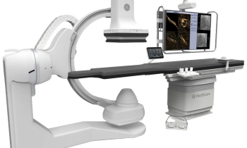
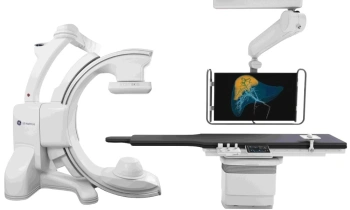
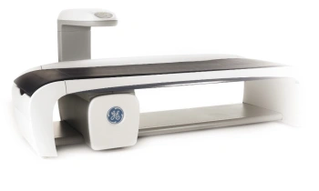
GE Signa and OR cradle
GE Healthcare and Barrow Neurological Institute at St. Joseph’s Hospital and Medical Center implementet a MR-guided intra-operative surgical suite. This new system includes one of the world’s most comprehensive portfolio of neuro-imaging solutions, which uses three-dimensional imaging to navigate and validate treatments to help increase both the accuracy and speed of interventions while improving patient outcomes.
The collaboration between GE Healthcare, Maquet GmbH & Co. KG and Barrow Neurological Institute at St. Joseph’s creates a MR surgical suite that offers multiple usage applications. In addition, both radiology and neuro-surgery clinicians collaborated on this project to help bring this unique technology to life.
The GE Neurosurgical suite provides MR imaging in one room of the surgical suite and an operating room (OR) in another. Surgery is performed on the Maquet MR/X-Ray compatible OR table system and real-time imaging is performed on the GE Signa HDx 3.0T MR scanner. Uniting the two enables patients to be moved quickly and safety from MR to OR – and vice versa – during surgical, minimally invasive, or therapy procedures.
“This new suite creates a variety of possible applications to better assist doctors with treatment and diagnosis, for optimum personalized healthcare,” said John Chiminski, vice president and general manager of GE Healthcare’s Global MR business. “Rather than waiting until after surgery to see whether a procedure went as planned, now patients can be evaluated during the surgical process to assess if the surgery is successful. A real-time 3.0T MR image can be evaluated without transferring the patient to a different table.”
Top-rated neurosurgeons say an important consequence of intra-operative image guidance will be the transformation of traditional brain surgery into image-guided surgery.
“Pre-operative imaging can detect and define tumors, but during surgery the anatomy can be distorted and the degree of tumor resection may be difficult to estimate,” said Dr. Robert Spetzler, neurosurgeon and medical director of Barrow Neurological Institute. “This technology will allow us to reevaluate and update our information allowing safer and more complete procedures. This improves our ability to gain maximum effectiveness while reducing the rate of complications. I am excited by the ability of utilizing our 3T MRI scanner in this manner to improve the outcomes of our patients.”
23.04.2007
More on the subject:More on companies: