Source: RSNA
News • Incidental findings identification
AI system for brain MRIs could boost workflows
An artificial intelligence (AI)-driven system that automatically combs through brain MRIs for abnormalities could speed care to those who need it most, according to a new study.
“There are an increasing number of MRIs that are performed, not only in the hospital but also for outpatients, so there is a real need to improve radiology workflow,” said study co-lead author Romane Gauriau, PhD, former machine learning scientist at Massachusetts General Hospital and Brigham and Women’s Hospital Center for Clinical Data Science in Boston. “One way of doing that is to automate some of the process and also help the radiologist prioritize the different exams.”
The research was published in the journal Radiology: Artificial Intelligence.
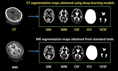
Dr. Gauriau, along with co-lead author Bernardo C. Bizzo, MD, PhD, and colleagues, and in partnership with Diagnosticos da America SA (DASA), a medical diagnostics company in Brazil, developed an automated system for classifying brain MRI scans as either “likely normal” or “likely abnormal.” The approach relies on a convolutional neural network (CNN). The researchers trained and validated the algorithm on three large datasets totaling more than 9,000 examinations collected from different institutions on two different continents.
Say you fell and hit your head, then went to the hospital and they ordered a brain MRI. This algorithm could detect if you have brain injury from the fall, but it may also detect an unexpected finding such as a brain tumor
Romane Gauriau
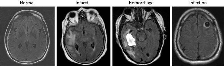
In preliminary testing, the model showed relatively good performance to differentiate likely normal or likely abnormal examinations. Testing on a validation dataset acquired at a different time period and from a different institution than the data used to train the algorithm highlighted the generalization capacity of the model. Such a system could be used as a triage tool, according to Dr. Gauriau, with the potential to improve radiology workflow. “The problem we are trying to tackle is very, very complex because there are a huge variety of abnormalities on MRI,” she said. “We showed that this model is promising enough to start evaluating if it can be used in a clinical environment.”
Similar models have been shown to significantly improve turnaround time for the identification of abnormalities in head CTs and chest X-rays. The new model has the potential to further benefit outpatient care by identifying incidental findings. “Say you fell and hit your head, then went to the hospital and they ordered a brain MRI,” Dr. Gauriau said. “This algorithm could detect if you have brain injury from the fall, but it may also detect an unexpected finding such as a brain tumor. Having that ability could really help improve patient care.”
The work was the first of its kind to leverage a large and clinically relevant dataset and use full volume MRI data to detect overall brain abnormality. The next steps in the research include evaluating the model’s clinical utility and potential value for radiologists. Researchers would also like to develop it beyond the binary outputs of “likely normal” or “likely abnormal.” “This way we could not only have binary results but maybe something to better characterize the types of findings, for instance, if the abnormality is more likely to be related to tumor or to inflammation,” Dr. Gauriau said. “It could also be very useful for educational purposes.”
Further evaluation is currently ongoing in a controlled clinical environment in Brazil with the research collaborators from DASA.
Source: Radiological Society of North America
22.04.2021