Computational neuroscience
The lost highway in the brain
A study three years ago sparked a medical mystery when it revealed a part of the brain not found in any present-day anatomy textbooks. Recently, Indiana University computational neuroscientist Franco Pestilli and an international research team published an article in the journal Cerebral Cortex that suggests this missing part of the brain may play an important role in how we understand the world -- despite getting "lost" for more than a century.
A long flat bundle of nerves called the vertical occipital fasciculus, or VOF, the structure appeared in textbooks on the brain for about 30 years around the end of the 19th century, then mysteriously dropped out of sight completely. Pestilli was also part of the team that first traced this missing brain structure to an 1881 publication by Carl Wernicke, a German-Austrian neuroanatomist.
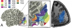
"Our new study shows that the VOF may provide the fundamental white matter connection between two parts of the visual system: that which identifies objects, words and faces and that which orients us in space," said Pestilli, senior author and assistant professor in the College of Arts and Sciences' Department of Psychological and Brain Sciences, whose work on the collaborative project began as a research associate at Stanford University.
"The structure forms a 'highway' between the lower, ventral part of the visual system, which processes the properties of faces, words and objects, and the upper, dorsal parietal regions, which orients attention to an object’s spatial location," he added. The first researcher on the paper was Hiromasa Takemura of the Center for Information and Neural Networks in Japan.
Based upon where the VOF terminates in the brain’s cortex, Pestilli said the VOF might play a critical role in the brain’s visual system, coordinating information transmission between brain areas important for "where we see" and "what we see."
In a long-standing debate, neuroscientists have argued the degree to which these two parts of the visual system function separately. The new study suggests that the regions work “hand-in-glove” to coordinate the body’s movement in relation to people and objects around it. There are "countless examples" that reflect how the two functions are intertwined, Pestilli said. Reaching for your keys, putting on your hat, driving to work each morning -- all require this what/where coordination. So does reading words on a page, which demands moving one’s eyes with respect to the words and letters.
What happens when this “what/where” coordination fails? Pestilli points to the title character in the late neurologist and writer Oliver Sacks’ “The Man Who Mistook His Wife for a Hat,” who, when asked to get his hat and put it on, tries to pick up his wife’s head to put it on his own.
In this strange disconnect between an object and its location, Sacks recognized the effect of a tumor or degeneration afflicting the patient’s visual system. The problem may have specifically affected the functioning of the VOF, Pestilli said, speculating on the symptoms of Sacks' patient.
But how did an entire anatomical structure in the brain suddenly go missing in the first place? The answer may be scientific rivalry. In their earlier paper, Pestilli and collaborators attributed the VOF’s disappearance to competing beliefs among 19th-century neuroanatomists. In contrast to Wernicke, Theodor Meynert, another prominent scientist in Germany, never accepted the new structure due to his belief that all white matter pathways ran horizontally. Over time, the VOF faded into obscurity.
In addition to contributing to the rediscovery and function of the VOF, Pestilli’s work is also helping map other parts of the human brain in unprecedented detail, using state-of-the-art neuroimaging and computational methods. This work will not only contribute to a deeper knowledge of the human brain -- such as how variations in brain structures relate to behavioral differences among individuals -- but will also strengthen the university’s other investigations into complex networks, including brain connectivity, primarily within the recently established IU Network Science Institute. "This new study really contributes to our understanding of how two important parts of the brain communicate," said Brian Wandell, a co-author at Stanford, "painting a picture of the brain that is highly interconnected."
Source: Indiana University
07.10.2015