Hitachi Aloka gives radiologists cutting edge in ultrasound
A Hitachi symposium demonstrated new high-end capabilities for ultrasound and reinforced the role of the radiologist in ultrasound examinations. "We are here not only to demonstrate capabilities, but to deliver a message," said Carlo Faletti, MD, from Turin, the chair for a symposium at the European Congress of Radiology focused on ultrasound multimodal fusion.
"We need to define the role of the radiologist in ultrasound examinations not only with high-quality scanners, but as high-quality operators," he said.
Following a compelling tour de force for the image fusion to improve prostate cancer detection, Thomas Fischer from Charité Hospital in Berlin echoed this message in a concluding comment. "The fusion of human brains on the medical team is as important as the images on new equipment," he said.
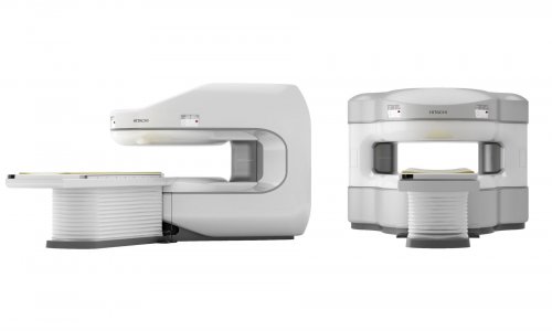
Once image volumes from MRI are loaded to the Hitachi HI VISION Preirus ultrasound scanner, images generated in real time from a trans-rectal ultrasound transducer can be viewed using advanced techniques including elastography, broadband Doppler and contrast harmonic imaging.
As he showed convincingly, the enhanced resolution of the B-mode alone yields a level of detail not seen before, enabling radiologists to detect, distinguish and closely examine tumors.
While prostate cancer was the immediate subject for his demonstration, the relevance of the multimodality fusion and high resolution ultrasound suggest interdisciplinary applications for nephrology, gynecology, gastroenterology and surgery, he said.
From head to foot, Taco Geertsma from the Hospital Gelderse Vallei in Ede, the Netherlands, demonstrated the clinical impact of ultra-high resolution technologies using the Hitachi Aloka ProSound F75 in musculoskeletal ultrasound examinations.
The detection and differentiation of smaller lesions with surprisingly short examination times increasing reveals the full extent of these previously unseen defects.
Visualization of nerves compressed by tendons for carpal tunnel syndrome is especially impressive with the new resolution levels, as well as subtle tendon abnormalities in the finger. The advanced visualization reinforces his role as a radiologist with surgeons.
"Any ultrasound system can show this ganglion cyst," he said. But using e-Flow signals and the high resolution, the patient's real condition is revealed to be nerve compression leading to a diagnosis of tarsal tunnel syndrome. By John Brosky.
05.03.2012