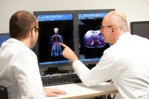
A sharper image with combined PET/MR technology
New instrument for cancer diagnostics provides precise images from inside the body
The German Cancer Research Center (Deutsches Krebsforschungszentrum, DKFZ) is sending a promising duo into the race against cancer: A new PET/MR system that can combine high-resolution images with functional information to improve cancer diagnosis.

The instrument also supports clinicians in choosing the best options for treatments and monitoring their progress.
Combining the methods of positron emission tomography (PET) and magnetic resonance imaging (MRI) makes it possible to simultaneously determine the location and size of a tumor while obtaining information about the metabolic activity of cancerous tissue. Radiologists obtain images with high spatial resolution and clear contrasts. The technology offers a key advantage over PET/CT, a method of combining PET with computer tomography (CT) that has been in use since 2000: MRI does not involve radiation exposure. In addition, PET/MR delivers better contrast in images of soft tissue. PET/MR is also superior when it comes to imaging organs in motion, such as the lungs. Professor Heinz-Peter Schlemmer, head of DKFZ's Radiology Division and a pioneer of the new combined imaging technology, sees "an enormous potential" in this advance in imaging technology and points out the opportunities it opens for personalized oncology: "We can image tissue structures much more sensitively than before. This enables us to better differentiate the tumor from healthy tissue and to characterize tumor biology more precisely." The additional information may help clinicians adjust therapies, such as radiation therapy, to the individual characteristics of a tumor. In the case of a tumor with a good prognosis, a patient may thus be spared the adverse side effects of aggressive therapy.
To date, only a few PET/MR instruments are in clinical use. Most of the 30-40 hybrid scanners that exist worldwide are used at research institutes and university hospitals in studies of their diagnostic potential. At DKFZ, radiologists and nuclear medicine specialists are using the combined imaging modality in clinical trials with, for example, prostate cancer patients. They are also studying the potential of PET/MR in patients suffering from multiple myeloma, lung cancer, brain cancer and malignant melanoma. Fundamental support for PET comes from the Clinical Cooperation Unit "Nuclear Medicine", led by Professor Uwe Haberkorn, and the Division of Radiopharmaceutical Chemistry. This unit was led successfully for many years by Professor Michael Eisenhut, who was succeeded by Professor Klaus Kopka on April 1, 2013. The nuclear medicine specialists are developing radiotracers, i.e. radioactively labeled biologically active substances that visualize the modified metabolism of cancer cells.
Radiologist Heinz-Peter Schlemmer is optimistic about the potential of the benefits of the new hybrid method, while noting that developments are still necessary. While working at Tübingen University Hospitals, Schlemmer witnessed the early development of PET/MR very closely. In November 2006, he published the first ever PET/MR images of the human body in the specialist journal "Radiology". Since the first prototypes of the hybrid instruments were used, there have been many advances in medical technology. "For many years now we have been working with Siemens Healthcare as a strong partner for further developing the technology," the radiologist said, and notes that the future will surely see many more improvements.
Schlemmer cannot tell whether PET and MR will be the "dream couple" for cancer diagnostics over the long term, because of important aspects of the technology that go beyond their medical benefits. There are technical challenges still to be resolved, and medical staff must be trained in the use of the complex systems. "And then we simply don't know yet whether the added value from the new technology will ultimately resist economic pressure in the healthcare system."
25.04.2013