SyMRI to be studied as a technology to characterize brain tumors
SyntheticMR and The University of Texas MD Anderson Cancer Center have signed a research agreement to study SyMRI to characterize brain tumors.
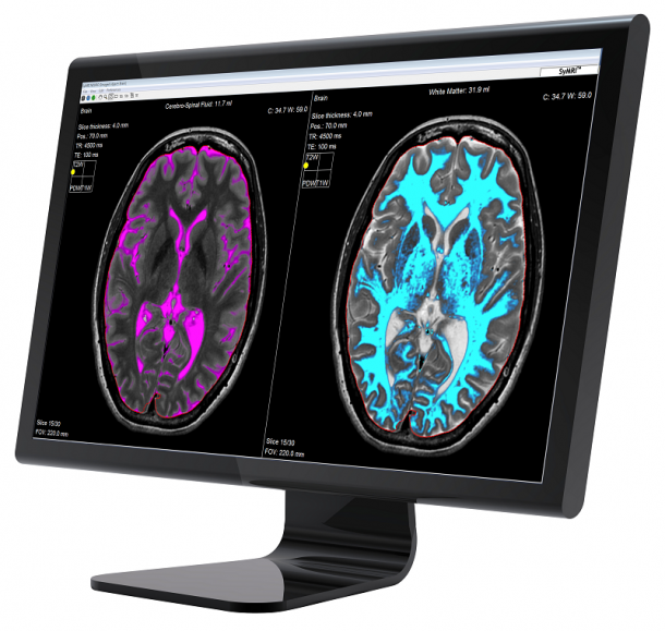
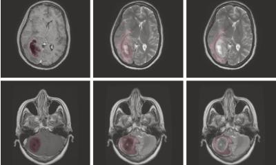
SyMRI provides quantitative information including tissue segmentation and T1, T2 and PD-maps, thus adding objective data to the MRI exam. The technology will be studied for its potential to add diagnostic value for various clinical applications including characterization of brain tumors.
SyMRI IMAGE generates multiple contrast images from a single 3 to 6 minutes scan, thus allowing a significant reduction in scan time compared to serial acquisition of each contrast image separately. Parameters are also adjustable post scan, enabling both fine tuning of images as well as recreation of additional contrasts. SyMRI NEURO adds quantitative data and automatic segmentation of brain tissue for efficient analysis and objective decision making. The agreement with MD Anderson includes a license of SyMRI IMAGE and SyMRI NEURO for research use at the hospital. SyMRI is CE-marked for clinical use in Europe but SyMRI is not available for clinical use in the USA.
Source: SyMRI
24.11.2015


