Article • Support for clinicians beyond initial diagnosis
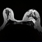
Enhancing breast imaging with AI
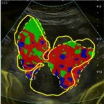
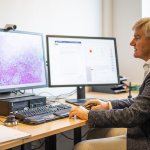
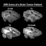
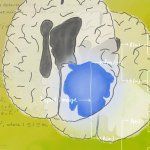
Artificial intelligence has a critical role to play in supporting clinicians beyond the initial breast cancer diagnosis. At the European Society of Breast Imaging (EUSOBI) annual scientific meeting in Aberdeen, Scotland, Professor Gerald Lip outlined how AI can enhance the performance of modalities such as ultrasound and MRI in supporting clinicians as they plan and deliver treatment for patients.