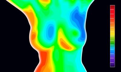
Image source: University of Aberdeen
Article • Support for clinicians beyond initial diagnosis
Enhancing breast imaging with AI
Artificial intelligence has a critical role to play in supporting clinicians beyond the initial breast cancer diagnosis. At the European Society of Breast Imaging (EUSOBI) annual scientific meeting in Aberdeen, Scotland, Professor Gerald Lip outlined how AI can enhance the performance of modalities such as ultrasound and MRI in supporting clinicians as they plan and deliver treatment for patients.
By Mark Nicholls
In his presentation, Professor Lip pointed to how AI can support lesion detection, characterisation and classification in ultrasound examinations. 'It also has a use in terms of reader education and improvement of specificity, but what is important in all AI implementations is the easy integration into workflow to support efficiency and adoption,' said the Clinical Director of the North East of Scotland Breast Screening Service.
He told delegates that AI research models with ultrasound are looking at tumour type prediction, prediction of neoadjuvant response, and prediction of the presence of axillary nodal metastases.
Reducing over-investigation in young women
An additional area of work in the UK is assessing whether AI can help avoid biopsy in ultrasound benign lesions in women under 35. 'We are actively looking at that in terms of where we can raise the age of not doing a biopsy for women with benign lesions because that is over-investigation and over-treatment, and this increases anxiety in a young female population,' Professor Lip explained.
In the realm of contrast-enhanced mammography (CEM), he said more data is needed but there is potential to use AI, particularly with synthetic contrast agents.
If we can enhance our technology with AI and not work in silos, we can work together to build a more patient centred and sophisticated future
Gerald Lip
It is with MRI that Professor Lip believes there are important opportunities with the integration of AI – such as with prediction of tumour response, risk assessment, breast cancer characterisation and segmentation tools based on radiomics. 'AI might be able to increase workflow and allow you to work more on complex and more interesting cases,' he told delegates. 'Again, they may also facilitate use of less contrast or even synthetic contrast agents.'
He noted that his colleagues at the University of Aberdeen are conducting research on Field Cycling MRI imaging – a new type of MRI developed in Aberdeen using low-fields of MRI to better characterise breast cancer. 'We are seeing good results with characterisation of DCIS (ductal carcinoma in situ) and of use AI algorithms in post-processing,' he said.
Pathology as the next growth area
While breast pathology may be some years behind radiology, Professor Lip suggested it could be the next big growth area for investors in tech. 'It definitely has areas that will help a lot more because that is where pharmaceutical and oncology companies are looking at classification tools so that we can segment tumours, do mitotic counting, and prediction for response. AI has clear benefits for breast pathology,' he noted, adding that Whole Slide Imaging (WSI) currently takes up significantly more data than standard radiology images.
Workflow tools and future considerations
Other aspects of AI that can support breast radiologists include voice technology, dictation tools, auto scribing and chatbots – all of which can help with workflow and time saving. Key considerations, he said, include post market surveillance, strategies for dealing with new hardware and software, procurement, data bias and diversity, and ethical concerns.
However, working with humans remains important in a world of GenAI and LLMs and the evolution of foundational AI and multi-mode models. 'By linking pathology, MRI and radiology together and putting in AI adaptors and tools, we are going to start talking about prediction and personalisation of treatments,' Professor Lip continued. 'If we can enhance our technology with AI and not work in silos, we can work together to build a more patient centred and sophisticated future.'
Profile:
Professor Gerald Lip is Clinical Director of the North East of Scotland Breast Screening Service and the Principal Investigator of the GEMINI prospective evaluation of mammography artificial intelligence. He is the current President of the British Society of Breast Radiology, and is the newly appointed Head of AI in Medicine at the University of Aberdeen Medical School, in line with his work in integrating artificial intelligence into healthcare.
04.03.2026