Image source: Beer et al., Urogenital 2020 (CC-BY 4.0)
News • CT & ultrasound fusion
‘Virtual biopsies’ could replace tissue biopsies in the future
A new advanced computing technique using routine medical scans to enable doctors to take fewer, more accurate tumour biopsies, has been developed by cancer researchers at the University of Cambridge.
This is an important step towards precision tissue sampling for cancer patients to help select the best treatment. In future the technique could even replace clinical biopsies with ‘virtual biopsies’, sparing patients invasive procedures. The research published in European Radiology shows that combining computed tomography (CT) scans with ultrasound images creates a visual guide for doctors to ensure they sample the full complexity of a tumour with fewer targeted biopsies.
Capturing the patchwork of different types of cancer cell within a tumour – known as tumour heterogeneity – is critical for selecting the best treatment because genetically-different cells may respond differently to treatment. Most cancer patients undergo one or several biopsies to confirm diagnosis and plan their treatment. But because this is an invasive clinical procedure, there is an urgent need to reduce the number of biopsies taken and to make sure biopsies accurately sample the genetically-different cells in the tumour, particularly for ovarian cancer patients.
High grade serous ovarian (HGSO) cancer, the most common type of ovarian cancer, is referred to as a ‘silent killer’ because early symptoms can be difficult to pick up. By the time the cancer is diagnosed, it is often at an advanced stage, and survival rates have not changed much over the last 20 years. But late diagnosis isn’t the only problem. HGSO tumours tend to have a high level of tumour heterogeneity and patients with more genetically-different patches of cancer cells tend to have a poorer response to treatment.
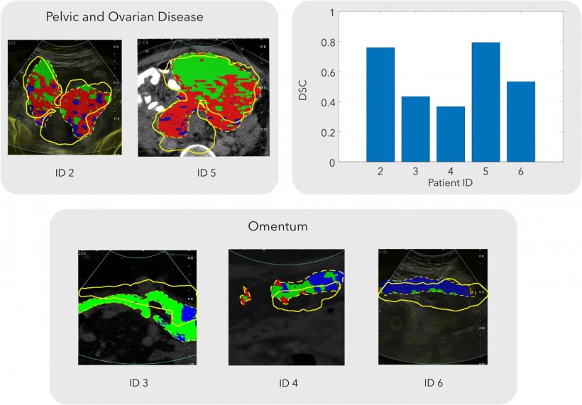
Image source: Beer et al., Urogenital 2020 (CC-BY 4.0)
Professor Evis Sala from the Department of Radiology, co-lead CRUK Cambridge Centre Advanced Cancer Imaging Programme, leads a multi-disciplinary team of radiologists, physicists, oncologists and computational scientists using innovative computing techniques to reveal tumour heterogeneity from standard medical images. This new study, led by Professor Sala, involved a small group of patients with advanced ovarian cancer who were due to have ultrasound-guided biopsies prior to starting chemotherapy.
This study provides an important milestone towards precision tissue sampling. We are truly pushing the boundaries in translating cutting edge research to routine clinical care
Evis Sala
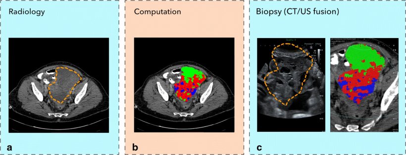
For the study, the patients first had a standard-of-care CT scan. A CT scanner uses x-rays and computing to create a 3D image of the tumour from multiple image ‘slices’ through the body. The researchers then used a process called radiomics – using high-powered computing methods to analyse and extract additional information from the data-rich images created by the CT scanner – to identify and map distinct areas and features of the tumour. The tumour map was then superimposed on the ultrasound image of the tumour and the combined image used to guide the biopsy procedure. By taking targeted biopsies using this method, the research team reported that the diversity of cancer cells within the tumour was successfully captured.
Co-first author Dr Lucian Beer, from the Department of Radiology and CRUK Cambridge Centre Ovarian Cancer Programme, said of the results: “Our study is a step forward to non-invasively unravel tumour heterogeneity by using standard-of-care CT-based radiomic tumour habitats for ultrasound-guided targeted biopsies.” Co-first author Paula Martin-Gonzalez, from the Cancer Research UK Cambridge Institute and CRUK Cambridge Centre Ovarian Cancer Programme, added: “We will now be applying this method in a larger clinical study.” Professor Sala said: “This study provides an important milestone towards precision tissue sampling. We are truly pushing the boundaries in translating cutting edge research to routine clinical care.”
Source: © University of Cambridge (CC-BY 4.0)
08.01.2021