Article • PRIMAGE project
Aiming AI at lethal paediatric tumours
La Fe University and Polytechnic Hospital in Valencia, Spain, is coordinating EU-funded program PRIMAGE, which uses precision information from medical imaging to advance knowledge of the most lethal paediatric tumours, by establishing their prognosis and expected treatment response using radiomics, imaging biomarkers and artificial intelligence (AI).
Report: Mélisande Rouger
About six months ago, the European Commission funded the PRIMAGE (short for PRedictive In-silico Multiscale Analytics to support cancer personalised diaGnosis and prognosis, Empowered by imaging biomarkers) project with over €10M, to help improve treatment and identify a tumour’s main characteristics without the need for biopsy, using computational processing of medical images on the cloud.

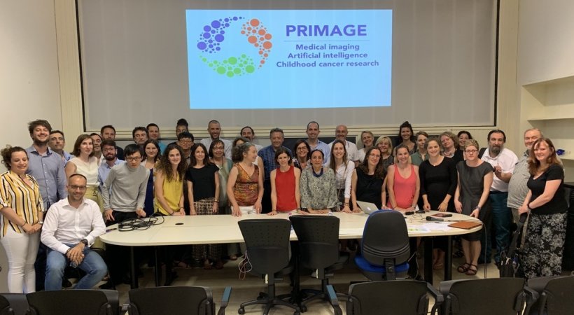
The PRIMAGE consortium will create a bank of images obtained through AI, using an open cloud-based platform to support decision-making in the clinical management of Neuroblastoma (NB), the most frequent solid cancer of early childhood, and Diffuse Intrinsic Pontine Glioma (DIPG), the leading cause of brain tumour-related death in children. The PRIMAGE platform will implement the latest advancement of in-silico imaging biomarkers and modeling of tumour growth towards a personalised diagnosis, prognosis and therapies follow-up. The ambitious project involves 17 European partners, including several internationally recognised institutions, and four leading industrial partners, all of which are working under the aegis of the Imaging Biomedical Research Group (GIBI230) at the La Fe Research Institute in Valencia.
The great value brought by PRIMAGE is that the study will use real world data as a foundation to help identify the best treatment for each individual patient, instead of collecting sample data, as in prospective trials. Exploiting the information from existing imaging biobanks and patient clinical files using advanced computational methods will help significantly to improve decision making in cancer management, according to GIBI 2030 Director Luis Martí-Bonmatí, Chairman of Radiology at La Fe Hospital.
Predicting disease right at the beginning
‘Using real world data in in-silico models can help establish imaging and molecular data’s capacity to estimate and predict right from the beginning of disease, to ultimately make the best therapeutic decision for each patient. This has, to our knowledge, never been done before for any type of cancer in a multicentric approach and using such disruptive technology,’ Martí-Bonmatí said. Being able to precisely estimate tumour phenotype, aggressiveness, stage and extension, define the best treatment plan and establish the most accurate prognosis will definitely help paediatric oncologists improve their decision-making, he added.
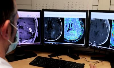
PRIMAGE will use the latest available technology in the field of computing and AI, for example machine learning software developed by QUIBIM, a spinoff of La Fe’s Research Institute that has received CE marks for its image post processing algorithms and is in charge for developing the PRIMAGE platform’s architecture, adaptation and design.
Recommended article

Article • Image analysis in radiology and pathology
"The time has come" for AI
AI has made an extraordinary qualitative jump, particularly in machine learning. This can help quantify imaging data to tremendously advance both pathology and radiology. At a recent meeting in Valencia, delegates glimpsed what quantitative tools can bring to medical imaging, as leading Spanish researcher Ángel Alberich-Bayarri from imaging biomarker company Quibim unveiled part of his work.
Knowledge acquired will benefit cancer treatment
Because there are so few reported cases, paediatric cancer tends to be forgotten by research
Luis Martí-Bonmatí
Cancer remains the first cause of non-traumatic death among children, but it has a very low incidence in this populations. Experts estimate that 500,000 EU citizens will be paediatric cancer survivors by 2020. Neuroblastoma is the most common extracranial tumour in children and represents 8-10% of all paediatric cancers. In Europe, 35,000 new cases are diagnosed each year, 1,000 in Spain alone.
Diffuse Intrinsic Pontine Glioma is a very rare disease in childhood and is associated with low survival (10%). There do exist palliative treatments and some research going on, but there is no curative treatment as yet.
PRIMAGE’s scope on these two diseases is expected to shed light on two of the less documented cancers. ‘Cancer is more rare in children than in adults. It is pertinent to use the data from international imaging biobanks in this setting, because we wouldn’t get many cases locally. Because there are so few reported cases, paediatric cancer tends to be forgotten by research, which makes it another interesting challenge for us, especially at La Fe Hospital, which is a European reference centre for neuroblastoma and DIPG,’ Martí-Bonmatí said.
Due to the peculiarities of computational approximation in these two types of tumours seen in childhood, investigation done in this area will also be applicable to other tumour types, to help advance research on cancer in general. ‘Our big interest is to give importance to real world data. If we’re successful, the tools and methodology used in PRIMAGE can be extrapolated to other cancers and improve knowledge and decision-making in this setting as well,’ Martí-Bonmatí concluded.
Profile:
Dr Luis Martí-Bonmatí is Chairman of Radiology and Director of the Medical Imaging Department at La Fe University and Polytechnic Hospital, Valencia. He is full member of the Spanish Royal National Academy of Medicine representing Radiology, and was founder and Director of the Research Group on Biomedical Imaging (GIBI230) within La Fe Health Research Institute. He also co-founded QUIBIM (Quantitative Imaging Biomarkers in Medicine), an innovative spin-off company from the Research Institute, although he no longer has a relationship with the company. He has been President of the European Society of Magnetic Resonance in Medicine and Biology (ESMRMB), Spanish Society of Radiology (SERAM), Spanish Society of Abdominal Radiology (SEDIA), and European Society of Gastrointestinal and Abdominal Radiology (ESGAR).
31.08.2019