Article • Experts outline European infrastructure
AI in health imaging: computational power isn’t everything
What will the future structure for artificial intelligence in health imaging across Europe look like? While the algorithms show great promise in collecting, storing, analysing, and using data to advance healthcare, delegates to a session on the topic at ECR 2023 in Vienna, also heard that it was important for the use of AI to move from research and more toward practical applications for patients.
By Mark Nicholls
Image source: Adobe Stock/Kingline
The session, entitled “Paving the way for a European infrastructure for AI for health imaging”, heard from speakers about projects currently under way, with a particular focus on cancer.
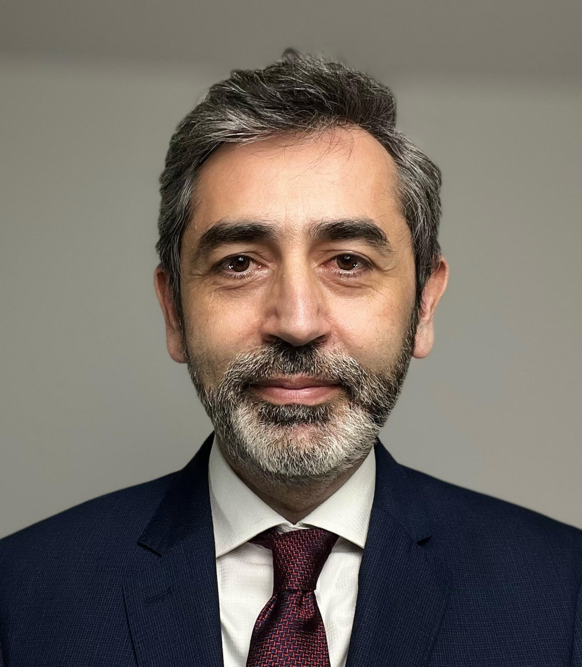
Dr Nikolaos Papanikolaou from the Champalimaud Research Foundation in Lisbon, Portugal, looked at the basic concepts of AI and radiomics, the challenges in a multi-centric setting, and on best practices to increase translation from research to clinic. He discussed the ProCancer-I project, an AI platform integrating imaging data and models, supporting precision care for prostate cancer. The major challenges in AI/radiomics include a lack of standardisation in image representation in the data and that radiomics features are highly sensitive to variations in image acquisition, he said.
While access to increased computational power is crucial, he also stressed the importance of the availability and accessibility to well curated high-quality data. He pointed out that medical images often non-harmonized, inconsistent, and heavily dependent on the way they have been acquired.
Intent on clinical translation
We need to prospectively, and consecutively, validate AI models into the clinical environment and organise randomised controlled clinical trials to document the impact on the clinical outcomes and to assess cost effectiveness
Nikolaos Papanikolaou
Papanikolaou said inconsistencies in feature extraction may deliver unreliable and non-reproducible results. This challenge should be addressed by applying harmonization methods, with AI models exposed to real world big data to learn invariant features. ‘But at the end of the day, the AI should not be only a research exercise – it should be developed towards an intention to be applied in the clinics,’ he said.
Areas of validity, usability and utility need to be ‘seriously addressed’ to convince people to start translating the technology into the clinic, he urged. ‘We need to prospectively, and consecutively, validate AI models into the clinical environment and organise randomised controlled clinical trials to document the impact on the clinical outcomes and to assess cost effectiveness.’
From fragmented to harmonized and structured data
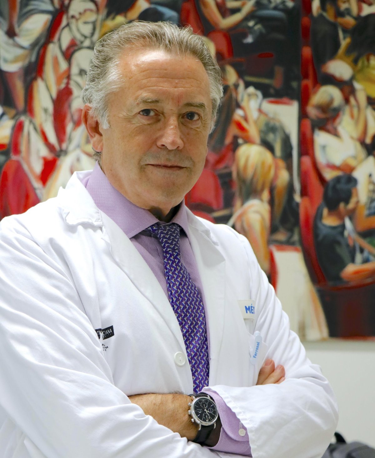
Image source: Hospital Universitari i Politècnic La Fe
Professor Luis Martí-Bonmatí, who co-chaired the session with Gabriel Krestin from Rotterdam, presented details and the goals of the EUCAIM (EUropean federation for CAncer IMages) project, which supports researchers to perform research on data. He looked at how fragmented health data silos can be linked and efforts made towards distributed big data harmonization and structure, while offering an appreciation of how federated learning can be used to construct clinical prediction models.
While there are opportunities for precision imaging and cancer, he also addressed the challenges: ‘Images are highly variable and data from Electronic Health Records is mainly non-structured and difficult to extract,’ said Martí-Bonmatí, who is EUCAIM’s Scientific Director.
‘We need a state-of-the-art platform for AI imaging to beat cancer but there is an innovation chasm,’ he said. To address this, EUCAIM aims to deploy a pan-European federated infrastructure to power up AI imaging to beat cancer; provide a research platform to develop and benchmark AI tools toward precision medicine; address the fragmentation of existing cancer imaging repositories; create a federated data warehouse approach to allow the construction of observational studies and populate a central repository; and allow the construction of observational studies. There is also the need to develop and foster standardization and harmonization tool via AI, the expert concluded.
Several other projects were also discussed during the session. Martin Starmans from the University Medical Center in Rotterdam, the Netherlands, discussed the “EuCanImage” project and looked at the role of the platform for streamlining European AI research in cancer imaging from data to deployment, with a focus on liver, colorectal and breast cancers ’to address real-world unmet clinical needs.’
With the CHAIMELEON project, Professor Ignacio Blanquer from the Technical University of Valencia, Spain, talked about the concept of in-situ access to data and examined the pros and cons of centralised repository models on harmonized images from patients with breast, lung, colorectal and prostate tumours.
Recommended article

Article • 'Chaimeleon' project
Removing data bias in cancer images through AI
A new EU-wide repository for health-related imaging data could boost development and marketing of AI tools for better cancer management. The open-source database will collect and harmonise images acquired from 40,000 patients, spanning different countries, modalities and equipment. This approach could eliminate one of the major bottlenecks in the clinical adoption of AI today: Data bias.
Delegates also were presented details on the INCISIVE project, which looks at ongoing research efforts for developing AI services for decision-support of clinical experts. Senior analyst and INCISIVE co-ordinator Gianna Tsakou underlined the importance of imaging and clinical data sharing, while acknowledging the benefits and hurdles of health data sharing and the role of clinical experts in this process.
In a further presentation, Angel Alberich-Bayarri from Quibim, Valencia, outlined the PRIMAGE project (HiE reported previously) with a specific focus on the architecture of an imaging platform for a paediatric cancer imaging biobank.
*The session was hosted by the European Institute for Biomedical Imaging Research (EIBIR), a non-profit organisation dedicated to the coordination of research in biomedical imaging and related discipline.
Profiles:
Dr Nikolaos Papanikolaou is a Principal Investigator in Oncologic Imaging at the Champalimaud Foundation in Lisbon, Portugal, and a research group leader of the Computational Clinical Imaging Group. He is also the Machine Learning Imaging Lead at the AI Hub of Royal Marsden Hospital in London, UK. He has published 110 scientific papers in peer-reviewed international journals and is the scientific coordinator of ProCancer-i, responsible for all AI activities of the consortium.
Professor Luis Martí-Bonmatí is the director of the Clinical Area of Medical Imaging Department at La Fe Polytechnic and University Hospital, Valencia, Spain. He is also the Scientific Director of the EUCAIM project.
29.06.2023