Article • A call for consistency at ECR 2026
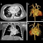
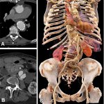
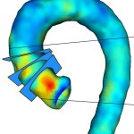
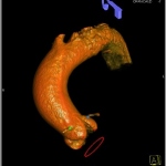
The aorta is an organ now – and radiology needs to catch up
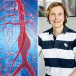
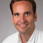
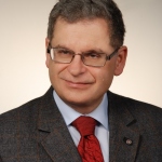
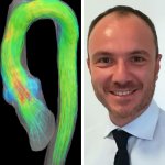
As the aorta gains recognition as a functioning organ rather than a simple blood conduit, the lack of standardised imaging protocols is becoming an increasingly pressing concern. At ECR 2026 in Vienna, Professor Nicola Galea from Sapienza University of Rome made a compelling case for unified imaging approaches, warning that inconsistency in measurement techniques can lead to flawed clinical…