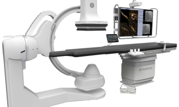
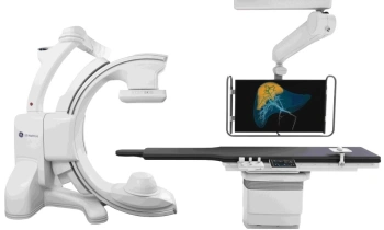
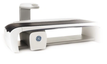
GE's new Discovery PET/CT 600 scanners go global
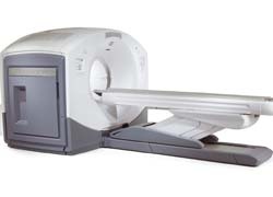
GE Healthcare's first Discovery PET/CT 600-series scanners are being installed in a number of leading clinics around the world.
"This first set of installations is a big step forward in the diagnosis and monitoring of disease", said Terri Bresenham, newly appointed vice- president and general manager of GE Healthcare's global Molecular Imaging business.
‘ ‘Partnering with clinics in the US, Europe and the Asia/Pacific region, we will be able to reach more patients globally with the latest advancements in oncology, neurology, and cardiovascular technologies.’
The new PET/CT platform, from GE Healthcare, was built for physicians and molecular imaging researchers, based on their need for more power to explore the potential of PET/CT imaging that includes better PET quantitative accuracy. GE points out that this new line of PET/CT scanners combine advanced molecular imaging tools with the large coverage low dose Volume CT helping to allow for earlier diagnosis, more accurate tumor location and better assessment of how a patient is responding to cancer treatment. Although mostly for use in oncology, PET/CT can also be used for assisting in the diagnosis of cardiovascular disease and neurological conditions, GE adds.
‘The new system leverages the high-speed, high-resolution capabilities of GE’s advanced CT, the LightSpeed VCT. Volume CT technology, with 40mm of coverage, dramatically reduces CT acquisition times and CT dose. Static organs can be imaged in as little as one second, the lung in as little as two seconds and a whole body trauma could be performed in less than 10 seconds,’ the manufacturer reports. ‘GE’s next generation of MotionFree imaging technologies are introduced in the new Discovery 600 series PET/CT, giving clinicians detailed images even in areas of the body subject to movement caused by respiration. GE’s breakthrough MotionFree imaging technologies are integrated with VUE Point FX, an advanced high-definition reconstruction technique. Combined, these technologies have the potential to improve small lesion detection, image quality, and better therapy response monitoring.’
Bichat University Hospital, in Paris, is among the first hospitals to receive a Discovery PET/CT 600-series scanner. Daniela Zimmermann of European Hospital, interviewed Professor Dominique Le Guludec, Head of the hospital’s Nuclear Medicine service, about its current use and early findings.
Initially, Prof Guludec explained, the scanner is mostly being used for oncology research, with about a third of its usage for the study of cardiovascular, infectious and inflammatory diseases. ‘For the time being, we don’t apply it much in neurology but will probably do so in the future,’ the professor explained. ‘We mostly carry out clinical research for new clinical indications, because although we know that most indications for PET-CT are justified, we have to prove it to our authorities for reimbursement of new indications.’ Within that need is having a certain number of cases but, equally important, she explained, is the provision of cost-efficiency evaluations. ‘The health insurers will only reimburse the scans if the clinical benefit is proven. We carry out the usual clinical studies in accepted indications, and pre-work in new indications.
‘In oncology, there are very established indications for PET-CT. We are looking primarily at ways to improve image quality through respiratory synchronisation. Our equipment is very sensitive. Because the scans are quicker we can do breathing synchronisation. A CT scan is very rapid. It only takes seconds, while PET image acquisition takes minutes. That means the patient has to breathe, which causes blurred images. To avoid blurring we use respiratory gating, which improves image quality as well as the quantification of the uptake of our tracers. We use mostly FDG, and we know that with FDG the uptake is higher when the tumor is more active. When you correct the uptake by respiratory gating, quantification is more accurate and you can evaluate the effect of the therapy. The PET-Volume-CT facilitates uptake quantification.’
Is motion a problem in PET-CT? ‘Motion is not a big problem for diagnosis but it might pose problems for accurate uptake quantifications because, when the diaphragm moves during respiration, the image you acquire is somewhat blurred. Consequently, quantification of the uptake in the tumor will not be completely accurate.
Is software support needed to improve quantification? ‘Yes, this is part of the Motion Free solution. Both the acquisition software that controls respiratory gating and the post-processing software need to support these efforts. We are working with General Electric Healthcare on further developing these solutions.’
How is PET-CT used for cardiovascular studies? ‘Recently, it has been shown that atherosclerotic plaques, which are at risk, are metabolically active and FDG can visualise these plaques in carotid arteries and the aorta. It is important to know the quantification of uptake in these plaques if the patient is at risk, or not, of cardiovascular events. In the USA, and now in Europe, there are some studies exploring vascular uptake of FDG. Also, we will soon work with rubidium, which means we will carry out perfusion studies with PET, which will be more accurate than SPECT in some patients. We already perform a lot of SPECT perfusion studies but we will use PET because, with PET, we can correct for attenuation, avoid some artefacts and also have an absolute quantification of the coronary reserve.’
What about soft and hard plaques? ‘Some plaques don’t affect the degree of stenosis that much, but they can cause a thrombosis and an acute event due to the thrombosis. We want to be able to differentiate between plaques that don’t pose a risk and those that do because they might cause thrombosis and consequently clinical events. Probably PET can help us to do that. Soft plaques are metabolically active because they have a lot of inflammatory cells. Inflammatory cells can catch the FDG and so you can obtain an image of the inflammatory characteristics of the plaque. You can distinguish between the two plaques. We also use hybrid imaging, which means we have a 64-slice CT linked to a PET. We can do the CT and the PET, so we see, live, precisely where the plaque is metabolically active. The CT gives us the calcium and anatomy of the vessel and we get information from both the PET and the CT images. So the images are matched to see function as well as morphology: the CT information (anatomy) and the PET information in the vessel (metabolic).
Is this not sufficient proof for reimbursement? ‘PET-CT findings have been correlated with histology, used in animal models and studied in patients who underwent carotid endarterectomy. However, for reimbursement purposes, we need to prove that PET-CT has a clinical impact on the prevention of events and thus on therapy costs.
How is PET-CT also being used for infectious and inflammatory diseases? ‘In our hospital, we see a lot of patients with infection, inflammatory diseases and diseases of the immune system. PET-CT is very well suited to evaluate these diseases. Be it retroperitoneal fibrosis, vascularitis or many immune diseases, we can visualise the complete organ involvement in one imaging process. As far as infectious diseases are concerned, we sometimes have patients with long-lasting fevers of unknown origin and the PET shows where the infection is located. In our clinical research we look for arguments to stop or continue therapy. For example, with tuberculosis every patient receives treatment for six months or one year, but some are okay before that and some need more. So we want to establish individual parameters which say this patient has no more bacteria and treatment can be stopped, while another patient still has bacteria and requires further treatment.’
Along with the Bichat University Hospital, the first Discovery PET/CT 600-series scanners are being installed at Miami Baptist Hospital (Florida), Queensland X-Ray at the Mater Hospital (Brisbane, Australia), Duke University Medical Centre (North Carolina, USA) and the Mayo Clinic (Minnesota, USA).
01.03.2009
- breast cancer (639)
- cardiovascular diseases (743)
- colon cancer (157)
- CT (617)
- imaging (1676)
- lung cancer (173)
- markets (547)
- PET/CT (192)
- prostate cancer (223)
- SPECT (31)