MR Angiography: A future without contrast media?
By Stefan G Ruehm MD PhD, Associate Professor of Radiology at the David Geffen School of Medicine, UCLA, California, and Director of Diagnostic Cardiovascular Imaging, CT, UCLA Radiological Sciences
Developments in MRI over the last few years have revolutionized the diagnosis and therapy of cardiovascular diseases. Contrast-enhanced MR angiography has established itself as a non-invasive, standard procedure for the diagnosis of vascular diseases in the thorax, abdomen and periphery.
[Bild-] It is characterized by fast acquisition times and lack of invasiveness. The three-dimensional display of data sets is similar to that achieved with conventional angiographic images and radiologists and clinicians are familiar with it. The paramagnetic contrast medium based on Gadolonium (Gd) is normally very well tolerated. Side effects such as allergic reactions are extremely rare. Unlike with iodine containing contrast media, if the maximum dose is adhered to there is no nephrotoxicity
However, the safe image of contrast-enhanced MR examinations has been questioned with recent reports about a link between the systemic and incurable disease nephrogenic systemic fibrosis (NSF) and the administration of contrast media containing Gadolinium. So far the occurrence of the disease appears to be limited to patients with severely limited kidney function. Ironically, it is often patients with increasingly deteriorating kidney function for whom an MR angiography is particularly indicated to eliminate a possible renal artery stenosis. In the past it was quite common for patients to be given twice or even triple the usual dose of contrast medium for this examination. Renal artery stenoses mostly develop because of arteriosclerotic changes. The systemic disease pattern of arteriosclerosis and associated diseases, such as high blood pressure and strokes, often require clarification in several vascular territories for the elimination of vascular pathologies. In the past, whole-body MR angiography was successfully used as an efficient screening procedure. All in all, it would appear sensible to reduce the total dose of contrast media because of the imminent risk of NSF. Whole-body MR angiography offers advantages here. It allows for more efficient use of the dose of contrast media administered compared with conventional protocols limited to a single vascular area. This makes it possible to reduce the amount of contrast media administered compared with the conventional procedure where separate doses of contrast media are injected for the different vascular territories to be displayed.
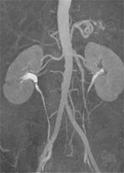
Recently published studies have proved that the use of the most up-to-date technology, particularly scanners with higher field strength (3-T), allows a significant reduction of the dose of contrast media required. Studies in our own institute confirmed that the use of the single dose Gd (0.1mmol/kg) definitely allows for combined MR angiography of the abdomen and the lower extremities without any sacrifices in quality (Pic. 1). In the past it was not uncommon to use double or even triple doses. Half an individual dose (0.05mmol/kg) appears diagnostically sufficient for the MR angiography of an individual vascular area (Pic. 2). A further reduction of the Gd dose could be achieved through the use of contrast media with higher relaxivity, such as binding of the Gd complex to albumin.
Despite promising strategies for the reduction of contrast media, examinations completely without contrast media would be desirable, particularly for patients with limited kidney function or for general risk and cost reduction purposes, such as screening protocols. Where in the past, and based on individual indication, native sequences such as Time of Flight (TOF) or PC MR angiography, which are still used today, were only of limited use as an alternative to contrast-enhanced MR angiography procedures because of motion artifacts, saturation effects and prohibitively long acquisition times for large vascular areas, state-free precession (SSFP) sequences are becoming increasingly important as an alternative.
Promising results have been achieved with Navigator SSFP sequences for the screening of high-grade renal artery stenosis, with sensitivities and specificities of 100% and 84% respectively. Our own research has shown that contrast media enhanced MR angiography definitely matches the diagnostic accuracy of SSFP sequences for the detection of pathologies of the aorta and thorax veins. Due to the ECG-synchronous data acquisition, native SSFP-MRI was actually superior to contrast-enhanced MR angiography for the display of the ascending aorta because of the lack of motion artifacts (Pic. 3). Although it is not yet clinically established, SSFP MR angiography currently appears to be the preferred procedure for the display of coronary arteries. However, it does have disadvantages compared with contrast-enhanced MR angiography for the display of smaller vessels. Moreover, the data acquisition times in thorax and abdomen, depending on the spatial resolution desired and other factors, such as breathing rhythm and cooperation of the patient, are often much longer. Additionally, SSFP MR angiography does not deliver functional information such as can be achieved with contrast-enhanced MR angiography with chronological data acquisition during injection of the contrast medium. Despite several limits of the sequence protocols for native MR angiography that are currently available, examinations entirely without Gd-containing contrast media appear to have advantages that must not be misconceived. It seems justified, dependent on the indication for patients with limited kidney function, to primarily use native MR angiography sequences and only to proceed to using contrast media enhanced sequences when the initial results appear unclear.
For patients with normal kidney function, we should weigh up the advantages of the potential gain of additional information resulting from contrast-enhanced examination protocols against the risk reduction achieved through foregoing the administration of contrast media, along with other aspects such as acceptance among patients and cost factors.
Contrast-enhanced MR angiography is sure to retain its outstanding significance in non-invasive vascular diagnostics in the future. However, in the interest of a general cost and risk reduction, careful assessment of individual indication along with selective use of protocols with reduced doses of contrast media or of protocols without contrast media is mandatory.
03.06.2008