The power of cardiac imaging and the invasive cardiologist
'The lynchpin for future success will be the unity of disciplines and intensive teamwork'
Progress in cardiac imaging diagnostics has made cardiac catheterisation less common. What may sound like 'fishing in foreign territory' is in reality the chance for interventional cardiologists to concentrate on, and specialise in, more innovative invasive procedures.

Dr Oliver Bruder, Senior Consultant in Cardiac Imaging at the Clinic for Cardiology and Angiology and Dr Christoph K Naber, Senior Consultant in Invasive Cardiology, both at the Elisabeth Hospital, Essen, both speakers* in the Cardiac Imaging 2008 Update session held during yesterday’s Medica Congress, consider this trend a win-win situation from which ultimately all patients will benefit.
In a European Hospital interview, Dr Naber said that the cases he now treats in the cardiac catheterisation laboratory are far more complex than just a few years ago. ‘There is a decreasing number of cases where I carry out a purely diagnostic clarification – this is mostly done by my imaging colleagues.’ According to Dr Naber there are currently three clinical relevant topics in interventional cardiology: aortic valve replacement via cardiac catheterisation, implementation of stents via the arteria radialis in the wrist and (cross-disciplinary) interventional vascular medicine. ‘Aortic valve replacement via catheter is of particular benefit to high risk surgical patients or those who previously could not even have surgery. This procedure entails the insertion of a “folded” heart valve through the femoral artery to the heart, where it either unfolds or is placed by a balloon, and then directly takes over the function of the diseased valve. This minimally invasive procedure significantly reduces patient recovery time.
‘Catheter access via the arteria radialis has the same advantages: The artery in the wrist is smaller, it is only punctured, significantly lowering the risk of bleeding. After the procedure, pressure is applied to the artery with a synthetic wrist band and the patient is immediately mobile.
‘The third area that interventional cardiology will increasingly cover is interventional vascular medicine. What is happening today is that experienced invasive cardiologists are performing examinations and treatments of the arteries of the legs, the cervical artery and of the kidney arteries. The training and experience required in this field, handling wires, balloons, guide catheters and so on, determines success or failure of the intervention. Of course, every vascular area has its unique features that have to be considered and mastered.
Asked (in jest) whether he feels a little ‘guilty’ that some medical colleagues must now find pastures new since a big portion of diagnosis has landed in the domain of non-invasive cardiac imaging, Dr Bruder’s reaction was immediate: ‘No, it’s great! Patients thank me for it. Of course non-invasive cardiology is pushing ahead with innovations such as those described by Dr Naber. As progress in imaging makes it possible to lower the number of cardiac catheterisations needed, the interventional side has to find new ‘adventures’. Non-invasive imaging considers itself a preparer – a service provider. With the aortic valve replacement, for instance, we deliver the decisive information and data so that catheters can be inserted precisely. So, we work hand in hand.
Imaging trends
Dr Bruder said their current work is mainly with MRI. ‘One of our joint objectives for the future is to carry out interventions that are currently only possible with modalities using radiation with MRI instead. Again, using the example of the cardiac valves, in the near future we hope to be able to fit these interventionally with the help of MRI, which is already done for some cases of the pulmonary valve diseases.
‘However, we are talking about developments within the next five to ten years. These MRI guided interventions need catheter material that is easily visible in the MRI. We are at an early stage: The material must be visible and compatible, i.e. ferrous materials pose a safety risk because of the magnet. This is a classic case of ‘safety & feasibility’.’
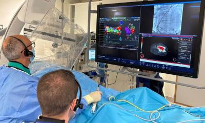
‘What is not actually a big issue these days is image fusion: We match MRI images with other procedures, such as electrophysiology. The electric map of the heart and the atrium is fused with MRI images, which is, for example, very helpful in the treatment of atrial fibrillation through ablation.
‘We expect enormous progress in non-invasive as well as interventional cardiology in coming years. A growing together of disciplines and intensive teamwork will be the lynchpin.’
* Dr Oliver Bruder, Essen:
Diagnosis of coronary heart disease with magnet resonance tomography
* PD Dr Christoph Naber, Essen:
Diseases of the aorta from the perspective of the interventional cardiologist
20.11.2008


