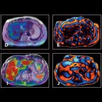
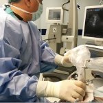
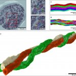
Search for: "virtual histology" - 33 articles found

Photo

Article • AI provides prognostic information

Next-generation deep learning models predict cancer survival

Deaths from cancer are currently estimated at 10 million each year worldwide. Conventional cancer staging systems aim to categorize patients into different groups with distinct outcomes. ‘However, even within a specific stage, there is often substantial variation in patient outcomes,’ Markus Plass, academic researcher from the Medical University of Graz, Austria, explained to Healthcare in…

Photo

Article • AI use in clinical diagnosis

Deep learning tool predicts tumour expression from whole slide images

A deep learning model to predict RNA-Seq expression of tumours from whole slide images was among the industry innovations outlined at the 7th Digital Pathology and AI Congress for Europe. Created by French-American start-up Owkin, the detail of how the company’s HE2RNA model provides virtual spatialization of gene expression was detailed to online delegates by senior translational scientist…

Photo

Article • Digital pathology

An exciting new era for tissue microarrays

A new generation of tissue microarrays are delivering more efficient and time-effective solutions to answering complex clinical and scientific questions. Sitting at the core of this new approach is digital pathology, allowing specific and targeted analysis of small areas of tissue.

Photo

Article • Imaging in tandem

MRE plus Fib-4 jointly detect liver fibrosis

Rather than using techniques separately, researchers have determined that coupling image-based and serum-based biomarkers achieves a higher diagnostic accuracy in detecting stage two liver fibrosis, or above. The study team, from the NAFLD Research Center, University of California at San Diego (UCSD), and colleagues at Yokohama City University in Japan, used magnetic resonance elastography (MRE)…

Photo

Article • Resecting brain tumours

Benefits of ultrasound in neurosurgical oncology

Italian neurosurgeon Professor Francesco Di Meco, explored the current and potential role of intra-operative ultrasound in neurosurgical oncology during the annual meeting of the European Association of Neurosurgical Societies (EANS) this October. The extent of resection is considered a prognostic factor in operative neuro oncology surgery and image-guided surgery is being regarded as one of the…

Photo

Sponsored • Histology/cytology equipment

Optimise pathology workflow with Hamamatsu whole slide imaging solutions

Hamamatsu Photonics understand the benefits and challenges of whole slide imaging systems. For over 15 years, we have been working to deliver robust and reliable scanners with optimal image quality. Our NanoZoomer whole slide scanner series quickly transform entire histology and cytology glass slides into diagnostic-quality digital images for duplication, annotation, storage, retrieval, and image…

Photo

Article • Need for modernisation

Digital pathology: Luxury or necessity?

The anatomical pathologist faces a crisis. Public and private labs suffer increasing caseloads, whilst pathologist numbers diminish for various reasons, including greater cancer prevalence associated with aging populations as well as improved cancer screening programs. Precision medicine typically involves more genetic testing and extensive use of immunohistochemistry to classify cancer and…

Photo

Article • Corona demographics

Why Covid-19 registries for cancer patients are so important

Due to compromised immune systems cancer patients are at higher risk of contracting infections. How does cancer impact on patients who also contract Covid-19? To collect this data, four cancer registries, one in the EU, one in the UK, two in the USA, have been established. The first large, multi-institution study of the impact of Covid-19 was conducted in Wuhan, China, and presented at the…

Photo

News • Diabetic damage

3D imaging shows how diabetes twists nerve fibers

In an international collaboration led by Lund University in Sweden, researchers have used synchrotron light to study what happens to the nerves in diabetes. The technique shows the 3D-structure of nerve fibers in very high resolution. “This knowledge can be used to map mechanisms for how nerve fibers atrophy and grow back. It means that we can better understand how diabetes affects the nerves…

Photo

Article • Heard at the British Cardiovascular Society conference

The role of nanomedicine in CV diagnosis

Nanomedicine will play an increasingly important role in future diagnosis and treatment of cardiovascular disease, a subject explored in detail by four expert speakers at the British Cardiovascular Society conference in Manchester in June. The conference heard that the technology – dealing with dimensions and tolerances of less than 100 nanometres, especially the manipulation of individual…

Photo

Article • Challenging process

Embracing digitised pathology

Investing in equipment and systems, training personnel and picking the right manufacturer to deliver a system that meets a hospital’s needs are key factors, according to Dr Peter Riegman, Head of Erasmus MC Tissue Bank at Erasmus MC in Rotterdam.

Photo

Sponsored • MALDI Tissuetyper

The rapifleX is now available as TOF/TOF

The MALDI Tissuetyper is a system that records spatially resolved mass spectra directly from tissue. This allows the direct measurement of proteins, lipids and other molecular classes without the need for antibodies or molecular probes. This results in highly multiplexed datasets in which hundreds or thousands of compounds are measured simultaneously.

Photo

Article • Moving on

I saw the future of pathology – and it’s digital

Healthcare is going digital. No doubt about it, Prof. Hufnagl predicts. Information and communication technologies have gone beyond moving data from one place to the other; they are triggering stellar improvements in healthcare: diagnoses are becoming ever more precise, therapies ever more personalised. The extent to which the individual clinical disciplines have progressed in their technological…

Photo

Article • Networking

Improvements in efficiency and quality through digitalisation

Increasing requirements for specialisation and diagnostic quality in pathology on the one hand and the importance of pathology findings for treatment planning on the other hand call for new solutions in pathomorphological diagnostics. One important starting point is the fast-paced opportunity for digitalisation along with communication systems which facilitate the storage and transfer of large…

Photo

Article • Vulnerable plaque imaging

Looking for the perfect modality

What's the ideal solution for vulnerable plaque imaging? 'A non-invasive imaging procedure with high spatial and temporal resolution, and without radiation exposure, and which provides information on coronary plaque composition precisely and in series.' Report: Axel Viola

Photo

40 years of CT scanning

Forty years ago an article was published that would change medical practice. In the British Journal of Radiology, English electrical engineer Godfrey N Hounsfield described how he had made a patient’s brain visible non-invasively by evaluating a large number of X-ray images of the skull taken from different directions.

Combinations advance endoscopic diagnosis and treatment

Endoscopy has advanced dramatically in the past decade with innovative technologies introduced by industry and novel procedures pioneered by physicians. Given a choice among the broad range of new tools, endoscopic surgeons simply want it all -- and are asking for more. During the Medica 2010 Congress, the Innovations in Endoscopy session rang with the word ‘combinations’.

Photo

Reading tissues

The trend towards personalised medicine implies the development of targeted cancer therapy. Tissue based examinations by pathologists play a key role in this trend. However, the relevance is still underestimated as pathologist Professor Manfred Dietel noted in his lecture at the European Forum on Oncology 2010 in Berlin, which explained what pathology already actually renders to targeted cancer…

Article • Reading tissues

Pathologists enable targeted cancer therapy

The trend towards personalised medicine implies the development of targeted cancer therapy. Tissue based examinations by pathologists play a key role in this trend. However, the relevance is still underestimated as pathologist Professor Manfred Dietel noted in his lecture at the European Forum on Oncology 2010 in Berlin, which explained what pathology already actually renders to targeted cancer…

Combining techniques advances endoscopic diagnosis and treatment

Endoscopy has advanced dramatically in the past 10 years with innovative technologies introduced by industry and novel procedures pioneers by physicians. Given a choice among the broad range of new tools and techniques, endoscopic surgeons have decided they want it all. And they are asking for more.

Photo

New perspectives in coronary plaque imaging using dual source CT

Ongoing technical developments in computed tomography (CT) such as dual source CT have established coronary CT angiography (cCTA) as a robust non-invasive imaging test for the assessment of coronary artery disease. The most important advantage of cCTA over conventional catheter-based coronary angiography is that not only the coronary lumen but also the entire coronary artery wall is visualized…

Photo

Arteries seen in a new light

New imaging technologies are opening a new chapter in interventional cardiology by offering something this widely practiced procedure has been missing -- vivid clinical evidence to assess effectiveness. Interventional cardiology has moved rapidly from opening blocked arteries by crushing plaque with inflatable balloons to reinforcing the walls of the re-opened arteries with flexible metal stents…

Radiologists are set to gain new control of images

CMIV has a new patent-pending technology that is set to radically change the way and speed at which radiologists work. The system could produce earlier diagnoses of certain disease, according to scientist Anders Persson MD PhD, Director and Member of the Board of the Centre for Medical Imaging Science and Visualisation (CMIV), at Linköping University, Sweden. CMIV has worked in close cooperation…

Subscribe to Newsletter