News • Diabetic damage
3D imaging shows how diabetes twists nerve fibers
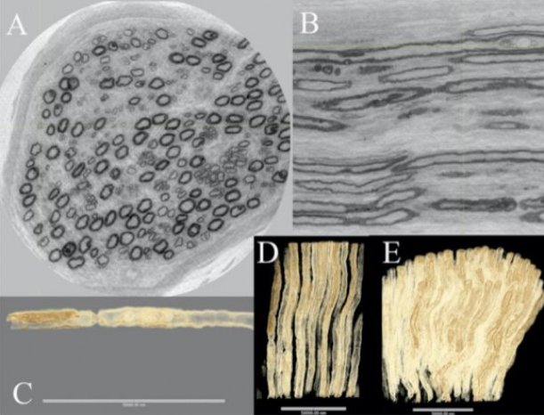
Image source: Lund University
In an international collaboration led by Lund University in Sweden, researchers have used synchrotron light to study what happens to the nerves in diabetes. The technique shows the 3D-structure of nerve fibers in very high resolution. “This knowledge can be used to map mechanisms for how nerve fibers atrophy and grow back. It means that we can better understand how diabetes affects the nerves in the arms and legs”, says Lars Dahlin, professor at Lund University and senior consultant at Skåne University Hospital.
The researchers published their findings in Scientific Reports.
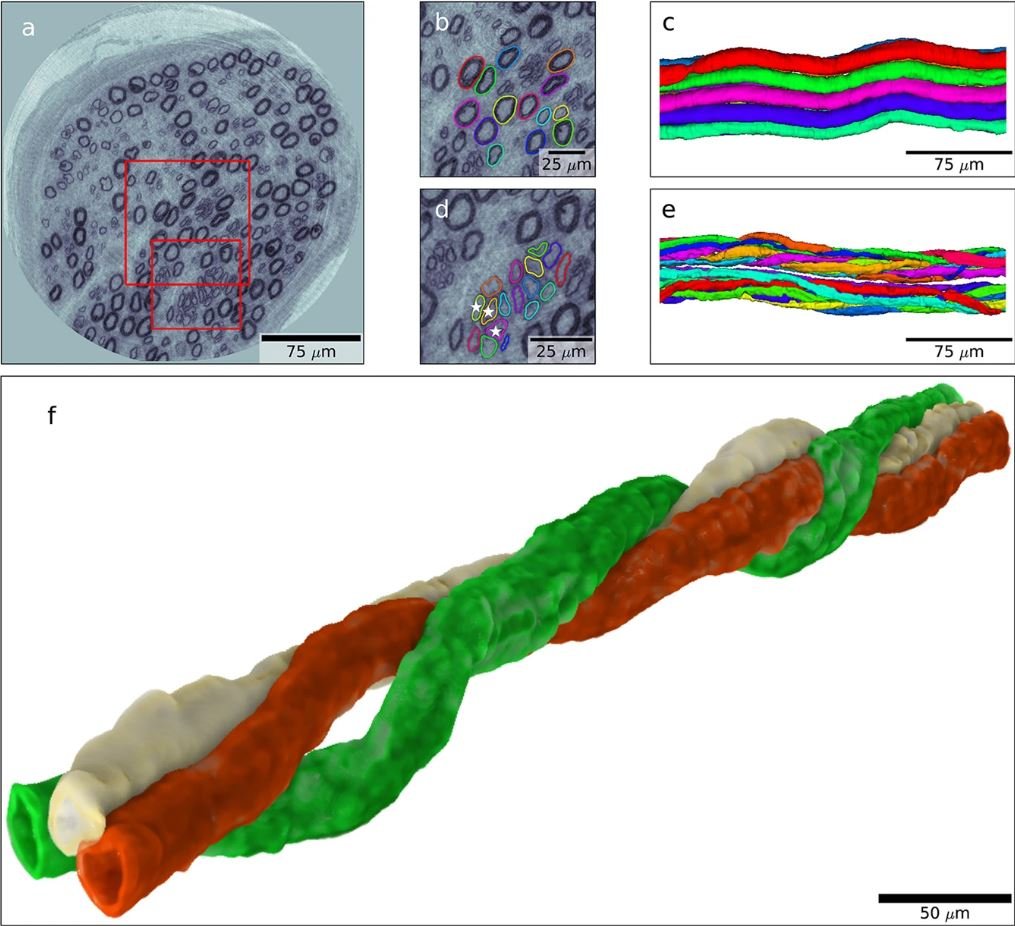
Image source: Dahlin et al., Scientific Reports 2020 (CC BY 4.0)
By using synchrotron light, the researchers have been able to show in detail what happens when nerve fibers in peripheral nerves are damaged. Such changes can occur in neuropathy, a nerve disease that affects patients with diabetes, but also in connection with surgical procedures. “In these cases, we know that nerve fibers atrophy. It appears that as they then grow, they take new paths - they are slightly more "confused". You could say they have poor GPS. But exactly what this looks like has not been shown before”, explains Lars Dahlin.
With previous techniques, it has only been possible to produce two-dimensional images. “This is a whole new way of studying nerves compared with histology, where you look at the tissue section by section in two dimensions. Here we get an image that allows us to rotate the nerve fiber and perceive details in a completely different way”, explains Martin Bech, medical radiation physicist at Lund University and one of the researchers behind the study.
This can deepen our knowledge of biological changes in diabetes, and in the long term alter treatment principles
Lars Dahlin
If you compare synchrotron light with the X-ray equipment used in a hospital, the synchrotron source is about a hundred billion times more intense. It is like a microscope, but with X-ray light that has a much shorter wavelength than regular light. This, in turn, allows you to study soft tissue at the cellular level without making incisions – referred to as virtual histology. In addition to researchers from Lund University and Skåne University Hospital, researchers at the European Synchrotron Radiation Facility (ESRF) in Grenoble, DTU in Copenhagen and Linköping University participated in the study.
The nerves that the researchers studied came from nerve biopsies from three individuals: one healthy person, a patient with type 1 diabetes and another one with type 2 diabetes. All of them had undergone surgery for carpal tunnel syndrome, a common condition, especially among those with diabetes. The researchers were able to map in detail what it looks like when, together with healthy nerve fibers, thin nerve fibers grow back and create something called regenerative clusters. They also found that when a nerve fiber is affected by diabetes neuropathy, it grows in a specific way. “The nerves grow back again in a spiral. Being able to see this in 3D gives us a unique opportunity to understand how nerve fibers grow, which is important both in diabetes neuropathy and in other direct damage to the nerves”, explains Lars Dahlin.
The researchers are now working on a larger follow-up study in which they hope to be able to further identify more nerve fibers. The study will investigate how the thickness of the nerve fibers varies, as well as the extent to which regenerative clusters occur. “This can deepen our knowledge of biological changes in diabetes, and in the long term alter treatment principles”, concludes Lars Dahlin.
Source: Lund University
06.05.2020