Combinations advance endoscopic diagnosis and treatment
Endoscopy has advanced dramatically in the past decade with innovative technologies introduced by industry and novel procedures pioneered by physicians. Given a choice among the broad range of new tools, endoscopic surgeons simply want it all -- and are asking for more. During the Medica 2010 Congress, the Innovations in Endoscopy session rang with the word ‘combinations’.
In his presentation on the diagnosis of early digestive tract tumours, Alexander Meining MD, from the Klinikum rechts der Isar, Munich, demonstrated how each new imaging technique developed by different manufacturers has been integrated into the hunt for neoplasia.
Magnification enhanced the traditional use of white light, then autofluorescence and various forms of computed virtual chromoendoscopy (CVC) enhanced mucosal surface contrast, much like using Photoshop on photographs.
Using a blue light to illuminate tissue, Olympus introduced narrow band imaging (NBI) which, he said, ‘… aids detection by the human eye and significantly improves the learning curve in endoscopy but is not necessarily better for detecting tumours.’
High powered magnification to over 400-fold endocystoscopy system (also from Olympus) further enhances examination of stained cells, but remains a surface examination.
More recently in vivo microscopy became possible using confocal laser scanning, a technology, Dr Meining said, which has proved effective in the early detection of tumours, adding that ‘virtual histology still has a lot of work to do before it can be adopted for routine clinical practice. ‘Again, it’s the combination of these techniques that allow us to go from a macro view to microscopic view and then, ideally, they will give us the confidence to perform a treatment for the condition we see in the same endoscopy session.’
'More importantly,' he pointed out, ‘these tools are teaching us what to look for and to look more closely.’
Presenting Progress in therapeutic endoscopy Pierre Deprez MD, Head of the Department of Gastroenterology at the Catholic University of Louvain, Belgium, said that, beyond the improved imaging, in recent years endoscopy has greatly benefited from the robust development of new tools for minimally invasive surgical interventions.
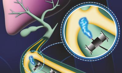
Combining techniques, such as endoscopic ultrasound with endoscopic retrograde cholangiopancreatography (ERCP) enables the diagnosis and treatment of conditions in the liver and gallbladder.
Crossing frontiers between endoscopy and surgery, innovations in natural orifice transluminal endoscopic surgery (NOTES) have introduced a toolbox of new instruments, including clips, stents, cutting tools and closure devices. ‘We used to be afraid of perforations during surgery,’ he said. ‘Now we routinely cut through lining to reach other organs.’ Endoscopy has also benefited from combining experiences by crossing borders, he added.
Techniques advanced by surgeons in Asia have shown that tumours can be fully removed in one piece, rather than cut into pieces -- the dominant Western technique.
Jean Pierre Charton MD, from the gastroenterology clinic of the Evangelisches Krankenhaus, Düsseldorf, pointed out that, from the pioneering pill cameras that first opened a view to the small bowel the technology has advanced to specialised capsules used to examine the colon and oesophagus.
Accepted for routine practice for certain indications, the pill camera presents an opportunity to encourage greater compliance to screenings for a significant patient population reluctant, or unwilling to undergo endoscopic exams.
29.12.2010


