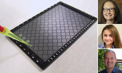
MALDI Tissuetyper
The rapifleX is now available as TOF/TOF
The MALDI Tissuetyper is a system that records spatially resolved mass spectra directly from tissue. This allows the direct measurement of proteins, lipids and other molecular classes without the need for antibodies or molecular probes. This results in highly multiplexed datasets in which hundreds or thousands of compounds are measured simultaneously.
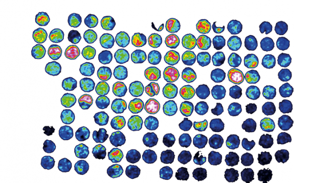
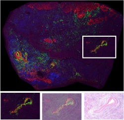
The acquired data can be used in different ways. For instance, using a classical biomarker approach, it is possible to compare different tissue or disease states to look for specific mass signals. Since the mass spectra can be also seen as reflecting complex tissue-specific phenotypes, it is also possible to use these patterns to develop bioinformatic tools that can then be used to classify unknown tissue. In the analysis of tumors, there can often be molecular differences in tissues that are histologically identical. The MALDI Tissuetyper can therefore be used to assist in the research of tumor heterogeneity. The system allows image co-registration; the ability to superimpose virtual microscopic slides with the MALDI data, enabling the detailed simultaneous analysis of histological and molecular information.
FFPE Tissue
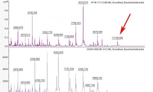
Most of the early development in mass spectrometry imaging has been conducted on frozen tissue, which allows for the analysis of intact proteins, lipids and endogenous metabolites. However, the interest in the analysis of formalin fixed paraffin embedded (FFPE) tissue has continuously increased due to the limited availability of frozen tissue and the wide availability of FFPE samples. Because of formalin crosslinking, intact proteins cannot be measured from FFPE tissue. Instead, spatially resolved tryptic digests are performed to measure tryptic peptides. While this adds additional sample preparation steps, it has the advantage that the mass spectrometry identification of tryptic peptides is easier than the identification of intact proteins.
The rapifleX
The introduction of the rapifleX MALDI Tissuetyper instrument last year was a milestone for mass spectrometry imaging. The system is a completely new design with the application of MALDI Tissuetyping in mind. The speed of MALDi imaging measurements was increased up to 20 fold, from a few pixels per second to up to 50 pixels per second. This now allows researchers to tackle problems that could not be targeted before, including the efficient measurement of large samples or many samples in research studies. This year, the scope of the rapifleX has been considerably widened by the introduction of the new rapifleX TOF/TOF. This instrument has a second ion source which allows MS/MS measurements to fragment and identify peptides and other compounds. In the context of the MALDI tissuetyper, this facilitates the identification of peptides as biomarkers or as part of classification patterns. Peptides can be identified by either direct measurement from tissue or more comprehensively by combining information from tissue imaging with information from LC-MS separations in the imageID workflow.
02.08.2016