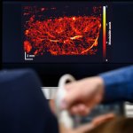
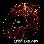
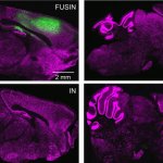
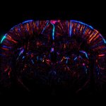
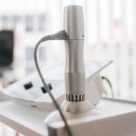
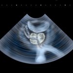
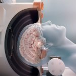
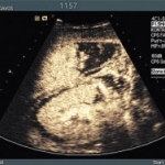
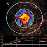
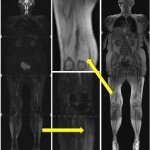
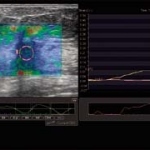
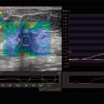
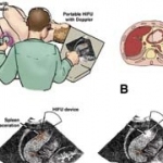
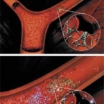
Search for: "Microbubble" - 46 articles found

Photo

Article • High-intensity focused ultrasound

HIFU ablation treatment for benign thyroid nodules

High-intensity focused ultrasound (HIFU) ablation is a noninvasively treatment for benign thyroid nodules that are causing distress to patients. Brian H. H. Lang, MD, Clinical Professor in the Department of Surgery at the University of Hong Kong, and chief of the Division of Endocrine Surgery at Queen Mary Hospital, is a preeminent investigator, proponent, and pioneer of this technique. He…

Photo

Article • Focused ultrasound

Histotripsy: fighting tumors with microbubbles

Focused ultrasound waves create microbubbles in a fluid – a phenomenon called cavitation. In a current study, this process is used to destroy liver tumors and metastases. In this Medica-tradefair.com interview, Prof. Maciej Pech talks about testing cavitation events generated during histotripsy, explains the process, and reveals its advantages.

Photo

Video • Enhanced therapeutic delivery

Focused ultrasound shows promise for Alzheimer's treatment support

A recent preclinical study from scientists at Sunnybrook Research Institute, Canadian Blood Services and the University of Toronto has demonstrated that focused ultrasound improves the delivery of intravenous immunoglobulin (IVIg), a blood product composed of antibodies from healthy donors, previously shown to have potential in treating a subgroup of patients with Alzheimer’s disease.

Photo

News • Reopening blood vessels

Microbubbles and ultrasound can help treat heart attacks

Doctors are using microbubbles and ultrasound to treat heart attacks – deploying these traditional diagnostic tools in an attempt to reopen tiny blood vessels, reduce scar size and restore heart function, according to a study described at a medical conference in Chicago. “This study shows for the first time that microbubbles can be used to both diagnose and treat small vessel obstructions…

Photo

Article • CEUS & the kidneys

Contrast-enhanced ultrasound for renal masses

Contrast enhanced ultrasound (CEUS) has proved in trials to be more accurate than computed tomography and MRI and can help eliminate the need for unnecessary biopsies and surgery. Dr Richard Barr, Professor of Radiology at Northeast Ohio Medical University, explains the benefits of the modality.

Photo

Article • Innovation

Siemens Healthineers tackle the problem of high-BMI patients with new Acuson Sequoia

Confidently scanning patients varying in size is one of the main difficulties in ultrasound imaging. With their new system Acuson Sequoia, Siemens Healthineers respond to this challenge. We spoke with Thomas Hartley, Senior Director of Global Marketing and Communication for Ultrasound at Siemens Healthineers, about the new device which aims to tackle some of the most pressing challenges in…

Photo

News • Groundbreaking technique

Noninvasive brain tumor biopsy on the horizon

Taking a biopsy of a brain tumor is a complicated and invasive surgical process, but a team of researchers at Washington University in St. Louis is developing a way that allows them to detect tumor biomarkers through a simple blood test. Hong Chen, a biomedical engineer, and Eric C. Leuthardt, MD, a neurosurgeon, led a team of engineers, physicians and researchers who have developed a…

Photo

Article • Diverse benefits

Experts present CEUS insights

In April 2016 CEUS received the USA’s FDA approval. This year‘s RSNA Samsung Symposium ‘Contrast-Enhanced Ultrasound (CEUS): Innovations and a Problem-Solving Tool in Clinical Practice’ provided an opportunity to take stock. For European Hospital, Professor André Clevert, Director of the Interdisciplinary Centre for Ultrasound at University Hospital Munich, Germany, describes the current…

Photo

News • Microbubbles

Bracco Imaging to innovate ultrasound for new personalized gene therapy

Bracco Imaging S.p.A., a global leader in diagnostic imaging, announced that it has initiated new experimental activities in its R&D Center in Geneva, Switzerland, to explore a new application for gas-filled microbubbles in the development of personalized gene therapy for treatment of chronic dysfunctional diseases related to lipid metabolism. Microbubbles have already revolutionized medical…

Photo

Article • Bubble business

A transducer halts abdominal bleeding

Stopping abdominal wall bleeding with contrast-enhanced ultrasound was just one of the exciting developments in CEUS presented at the Bubble Conference 2017 in Chicago. When you cut your finger you apply pressure to the wound until the bleeding stops. Professor Dirk-André Clevert from the Institute for Clinical Radiology at the Ludwig Maximilian University Hospital, Munich, Germany, remembered…

Photo

News • Contrast agents

SonoVue receives EU approval for detection of vesicoureteral reflux

Bracco Imaging S.p.A., a company in the diagnostic imaging business, announced today that its contrast agent SonoVue (sulphur hexafluoride microbubbles) is the first ultrasound contrast agent to obtain approval in the European Union for its use in ultrasonography of the urinary tract for the evaluation of suspected or known vesicoureteral reflux in pediatric patients.

Photo

Article • Contrast enhancement

Sonic boom with bubbles

Illuminating blood vessels, opening the blood-brain barrier and delivering drugs. What will be the next big thing that tiny microbubbles can do?

Photo

Article • Therapy

Molecular imaging mines deeper

The view across the Atlantic – it fills Professor Fabian Kiessling, Chair of Experimental Molecular Imaging at the RWTH Aachen (Rhine-Westphalia Institute of Technology Aachen), with optimism. The USA offers more opportunities for molecular imaging. Only recently, new tracers for Alzheimer’s were accepted as reimbursable in some centres, whilst the development of new diagnostics in Europe…

Photo

The complex science behind microbubbles

At Bracco Suisse SA in Geneva all efforts are dedicated to contrast media for ultrasound scans. During their visit to the firm’s research centre and manufacturing site, Daniela Zimmermann and Ralf Mateblowski met with François Tranquart MD PhD, general manager of the Bracco Suisse research centre, to hear why SonoVue is now Europe’s most popular ultrasound contrast agent, with research…

Photo

Bringing contrast into play

Although many new features in US-guided interventions are being marketed, which are really necessary, which just nice-to-have? It’s a question to be faced by experts during the refresher course ‘Interventional ultrasound’ at WFUMB 2011. One of the most established ultrasound techniques in minimally invasive procedures is contrast enhanced ultrasound (CEUS) – a tool that is safe, gentle…

Photo

Microbubbles in tumour therapy

Contrast agents have opened up entirely new possibilities are taking shape for ultrasound, above all in oncology. Following the publication of guidelines on the clinical use of contrast-enhanced ultrasound (CEUS) by the European Federation of Societies for Ultrasound in Medicine and Biology (EFSUMB) in 2004 and 2008, at this year’s World Ultrasound Congress, WFUMB and EFSUMB will present joint…

Contrast enhanced tumour studies

Medical imaging has recently advanced so rapidly that it should halt. Applying more power to computed tomography (CT) and magnetic resonance imaging (MRI) scanners is becoming too dangerous for patients and healthcare workers. Magnets for the next-generation MRIs are so powerful that they must be moved to a separate building on hospital campuses, while CT radiation levels have risen to alarming…

Photo

Modern radiological diagnostics for the evaluation of muscle diseases

Muscular diseases belong to a heterogeneous group with various causes like neurogenic, metabolic, dystrophic, or inflammatory mechanisms as well as channelopathies leading to disorders of the muscle cell membrane potential. In most progressive disease cases the result is a focal or general muscle weakness that, unfortunately, is a very unspecific symptom. Standard neuromuscular literature…

Photo

Royal Philips Electronics leads Sonodrugs Project

Philips leads the €15.9 million project to develop image-guided localized drug delivery technologies that could significantly impact the treatment of cancer and cardiovascular disease. The goal is to maximize therapeutic efficiency and minimize side effects of treatments for cancer and cardiovascular disease. The hope is that this level of control will also provide a means of tailoring the…

Portable real-time 3-D ultrasound for brain scans

USA - 3-D ultrasound technology developed at Duke University provides images of the brain vessels in real-time, which could be miniaturised in coming years for use in ambulances. In an initial pilot study, the system has passed the proof-of-principle.

Photo

The brain in three dimensions

Stroke treatment is a question of time. The faster the cerebral infarct can be diagnosed, the less the brain will be damaged. But unfortunately it is not always easy to get the patient to a brain scanner within the required three-hour window. Real-time 3D ultrasound might bring the solution, according to researchers from the Duke University in Durham, NC.

Photo

Trauma care

By Shahram Vaezy, Associate Professor of Bioengineering at the University of Washington, Washington, Seattle, and Vesna Zderic, Assistant Professor at Electronic and Computer Engineering Department at George Washington University, Washington DC.

Photo

Magic Microbubbles

The introduction of a new generation of microbubbles has created opportunities for ultrasound, and this is particularly true in the liver, where study of the arterial, venous and sinusoidal phases with an agent such as SonoVue at low ultrasound power levels (Iow mechanical index, MI) provides unique information. Similar diagnostic advantages can be obtained in diseases of the kidneys, spleen and…

Photo

Application of Vascular Recognition Imaging (VRI)

Vascular Recognition Imaging (VRI) is a low MI broadband colour Doppler method that images the interactions of Sonovue with microbubbles in a non-destructive manner as perfusion images of a quality never before achieved. The perfusion image represents the low velocities of unmoving or barely moving microbubbles as green-coded tissue perfusion, while the higher velocities of the veins and arteries…

Photo

Contrast Harmonic Imaging at its best

Over the last years Toshiba has actively participated in the development of Contrast Harmonic Imaging. Around 1996 one of the first agents that became available on the European market was Levovist TM (Schering AG, Berlin, Germany). This was a galactose-based contrast agent. Levovist could be used as a Doppler signal enhancer throughout the entire vascular system. With the development of these…

Photo

Contrast agents in Sonography

Ultrasound contrast agents (USCA) or echo enhancers consist of minute gas containing microbubbles that have a high reflectivity when exposed to an ultrasound field. The history of USCA started in the mid of the 1960s. The cardiologist Joyner performed echo cardiography during the injection of saline solution into the aortic root via a catheter. During the injection he observed bright signals on…

Subscribe to Newsletter