The brain in three dimensions
Stroke treatment is a question of time. The faster the cerebral infarct can be diagnosed, the less the brain will be damaged. But unfortunately it is not always easy to get the patient to a brain scanner within the required three-hour window. Real-time 3D ultrasound might bring the solution, according to researchers from the Duke University in Durham, NC.
Real-time 3D ultrasound might provide an imaging tool for stroke.
After a stroke the first three hours are crucial for further life. Brain damage of patients who are diagnosed and treated with the vital re-perfusion therapy in this time, will be limited and may offer them a life without being handicapped. But physicians know how difficult it is, getting patients to a brain imaging scanner within this time and to look for the vessels.
In the future real-time 3D ultrasound might provide a solution. Researchers from Duke University in Durham, NC, investigated the potential of this technique for cerebrovascular imaging and reason that with further refinements the system can be a truly portable, brain-imaging tool that will be suitable for screening. Patients with stroke-like symptoms can than be assessed at once, and physicians alerted if urgent re-perfusion therapy is required.
“I think it’s safe to say that within five to ten years, the technology will be miniaturized to the point where emergency medical technicians in an ambulance can scan the brain of a stroke patient and transmit the result ahead to the hospital”, says Stephen Smith, professor of bioengineering at Duke, who led the research.
Unlike previous reports of cerebrovascular imaging with ultrasound, in which data were reconstructed offline, the method deployed at Duke produces 3D data in real time. The system uses a 2D matrix phased array to scan a 65°-90° pyramid, producing up to 30 volumes per second.
"Being real-time is one of the major advantages of the technique," said graduate student Nikolas Ivancevich, first author of the paper. "There is instant 3D feedback and monitoring for the treatment of cerebrovascular disease."
Through the skull
The researchers have also developed a way to compensate for variations in skull thickness across the face of the ultrasound transducer. Left uncorrected, this variance can degrade image resolution and contrast.
The image correction method devised at Duke currently requires the ultrasound transducer to be held motionless for 20 seconds whilst data on the passage of echoes through bone are processed. A fixation device is used to prevent even the slightest movement. "If the technique proves useful, future systems could reduce the time required to less than a second," Ivancevich noted.
A pilot trial of the real-time 3D technique was carried out on 17 healthy volunteers. Doppler imaging was performed before and after injection of a microbubble contrast agent. All subjects were imaged with the ultrasound transducer placed on each temple of the head where the skull is thinnest (temporal window). Nine of the subjects were also examined with the transducer pointing upwards from the base of the neck (suboccipital window).
The results were reviewed independently by a vascular sonographer and a neurologist. Contrast was required to visualise blood flow in all cases. Both observers detected the major ipsilateral blood vessels in 71% of study subjects when imaging via a temporal window, and the complete circle of Willis (ipsilateral and contralateral vessels) in 51% of cases. They also both detected the entire vertebrobasilar circulation in 22% of subjects imaged from the suboccipital window, and the basilar artery in 44%.
Phase aberration correction was performed on just one volunteer as a proof-of-principle. Both observers agreed that the corrected 3D volume showed an extra, contralateral blood vessel that could not be seen in the uncorrected volume.
Team members now want to test their in vivo image correction technique on more volunteers. This should help determine the level of diagnostic value that is added to transcranial ultrasound. Additional studies are planned to assess the sensitivity and specificity of contrast-enhanced, real-time 3D transcranial ultrasound compared with either digital subtraction angiography or MR angiography.
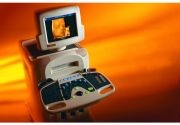
Photo: Philips Healthcare
25.06.2008
More on the subject: