US technique identifies subtle evidence of early PAD
An ultrasound imaging technique that picks up early evidence of peripheral arterial disease (PAD) currently missed in conventional tests has been developed by researchers* at Oregon Health and Science University (OHSU).
PAD frequently goes undiagnosed in its early to mid stages, partly due to the lack of cost-effective diagnostic tools that can detect the disease before it has become serious. If approved for clinical use this new technique could lead to early treatments that would prevent serious complications.
Lead investigator Jonathan R Lindner MD, Professor of Medicine (cardiovascular) at the OHSU School of Medicine, and his associates, devised the test during a study involving 26 control subjects with no history of coronary disease, hypertension or diabetes, and 39 patients with symptomatic PAD, 19 of them type 2 diabetics.
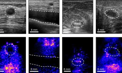
Lipid-shelled microbubbles were injected intravenously as an ultrasound contrast agent to evaluate microvascular blood flow in the calf muscles of subjects’ legs at rest and during exercise. These microbubbles behave and circulate in much the way red blood vessels do and they ‘ring’ in an ultrasound field giving off a strong signal. The results from blood flow imaging were compared with those from the most commonly used non-invasive diagnostic tests.
Contrast-enhanced ultrasound imaging outperformed most conventional forms of diagnostics in measuring and evaluating impairment in a patient’s ability to recruit blood flow in the legs during modest exercise, he reported. Specifically, the study found that patients with PAD had lower microvascular blood flow in their calf muscles than in the control subjects after two minutes of plantar-flexion exercise.
‘We don’t have good diagnostics for PAD, Prof Lindner pointed out, ‘partly because a lot of methods are based on measuring what’s going on in the big vessels, arteries and veins. PAD is a complex, very diffuse disease, which often involves functional abnormalities in the microcirculation system, the tiniest small vessels that go into muscle, bone, skin and connective tissues. How well the microcirculation system is functioning is what determines how well tissue is getting fed, which is the critical issue.’
Current PAD diagnoses rely mainly on detection of abnormal pulse volumes in the lower limbs, or blood pressure differences between separate readings at the ankle and arm (ankle-brachial index), but these are only effective in detecting moderate-to-severe cases and do not always detect disease in small vessels.
‘The real benefit of a test like this – which only takes about five minutes and doesn’t require anything beyond the equipment and capabilities already in place in most vascular laboratories – is their value for selecting the right therapies,’ the professor explained. ‘There are new drugs in clinical trials at OHSU and elsewhere that involve gene and stem cell therapies aimed at improving the circulation by promoting new blood vessel growth. But the symptomatic benefits you get from these therapies generally can’t be evaluated by conventional tests because they don’t really test tissue perfusion. If you want to look at the improvement in perfusion, why not look at perfusion?’
Further studies are needed on people for whom no diagnosis has yet been made. The microbubbles used are already approved for human use, although the PAD test is currently an off-label application.
Additional training in how to receive the microbubble signals is necessary, Prof Lindner pointed out, but there are few other hurdles before the test can be used in vascular clinics in general.
* Study findings: JACC: Cardiovascular Imaging, a peer-reviewed journal of the American College of Cardiology Foundation. [J Am Coll Cardiol Img, 2008; 1:343-350, doi:10.1016/j.jcmg.2008.04.001] http://imaging.onlinejacc.org/cgi/content/abstract/1/3/343.
19.11.2008